Blog

NEWS AND DENTAL EDUCATION

We share informative articles and news.


20/Feb/2011

What is root canal treatment

Root canal treatment on front teeth

Root canal treatment or sometimes refers as RCT is a treatment used to repair and save a tooth that is badly decayed or becomes infected. During a root canal procedure, the nerve and pulp are removed and the inside of the tooth is cleaned and sealed. Without treatment, the tissue surrounding the tooth will become infected and abscesses may form (below).

Gum abscess

What is root canal?


Root canal is the term used to describe the natural cavity within the center of the tooth. The pulp or pulp chamber is the soft area within the root canal. The tooth’s nerve lies within the root canal.
A tooth’s nerve is not vitally important to a tooth’s health and function after the tooth has emerged through the gums. Its only function is sensory — to provide the sensation of hot or cold. The presence or absence of a nerve will not affect the day-to-day functioning of the tooth.

What Damages a Tooth’s Nerve and Pulp in the First Place?

Big decay

A tooth’s nerve and pulp can become irritated, inflamed, and infected due to:
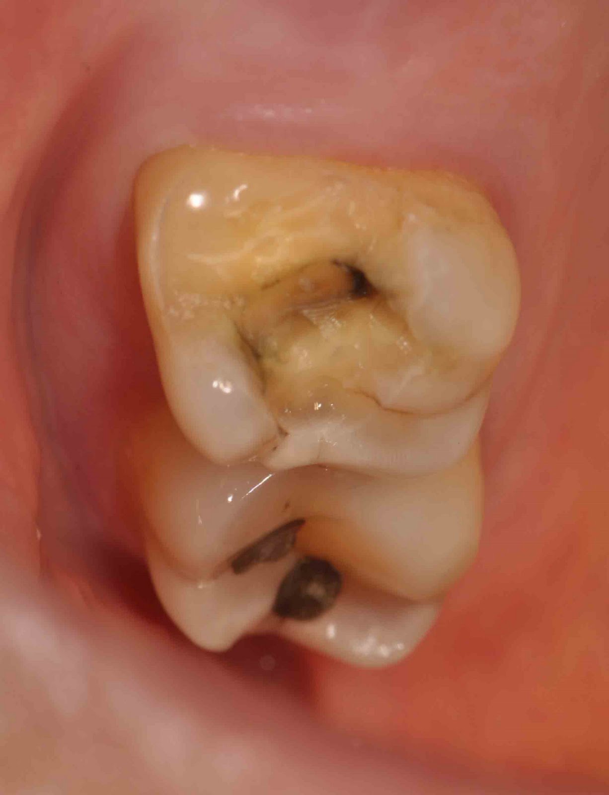
  • deep decay
  • repeated dental procedures on a tooth and/or large fillings
  • a crack or chip in the tooth
  • or trauma to the face.

How does dentist know whether the tooth needs RCT?

There are several symptoms that usually lead dentist to conclude that the tooth requires RCT:

Pain
Tooth Discolouration


  • Pain which is severe and prolong that affect  daily activities
  • Throbbing pain that usually disturb sleep at night
  • Feel painful when biting on the affected side
  • Prolonged sensitivity/pain to heat or cold temperatures (after the hot or cold has been removed)
  • Discoloration (a darkening) of the tooth
  • Swelling and tenderness in the nearby gums
  • A persistent or recurring pimple on the gums (gum abscess)

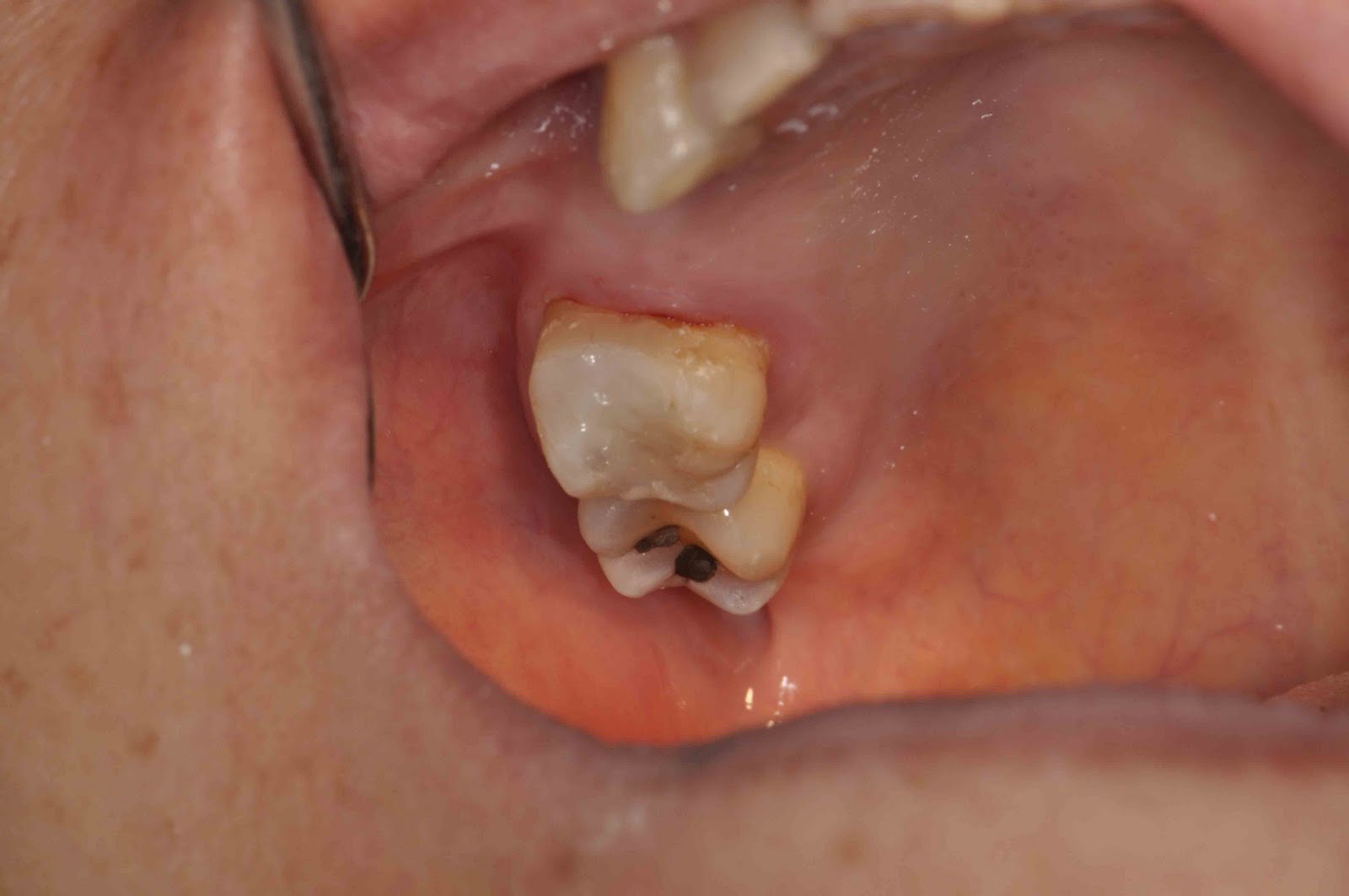
From dental examination:

Pulp testing
  • A very large cavity or deep restoration on that tooth
  • Feels very painful on percussion on that tooth
  • Not responsive to the vitality test using a Pulp Tester
  • A radiolucency lesion over the tip of the root on radiograpghic examination

How does Root Canal Treatment done?

Step-by-step of root canal treatment

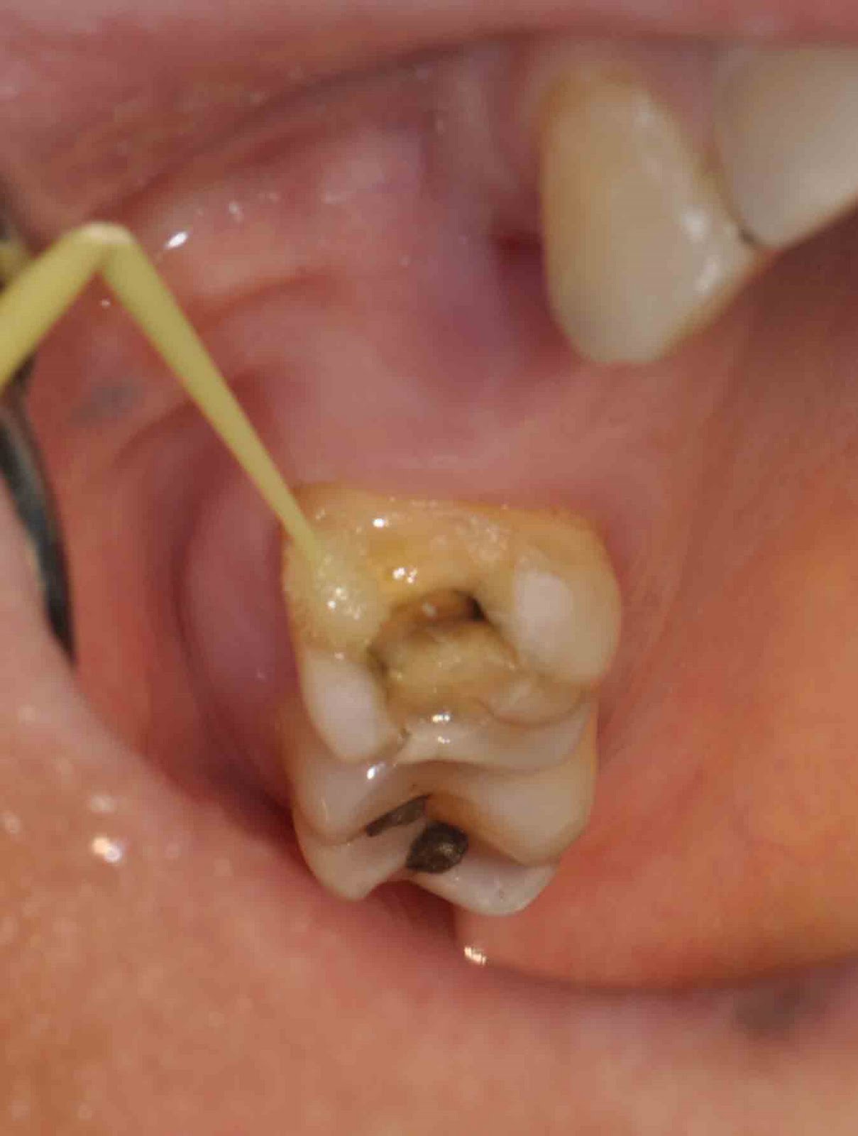
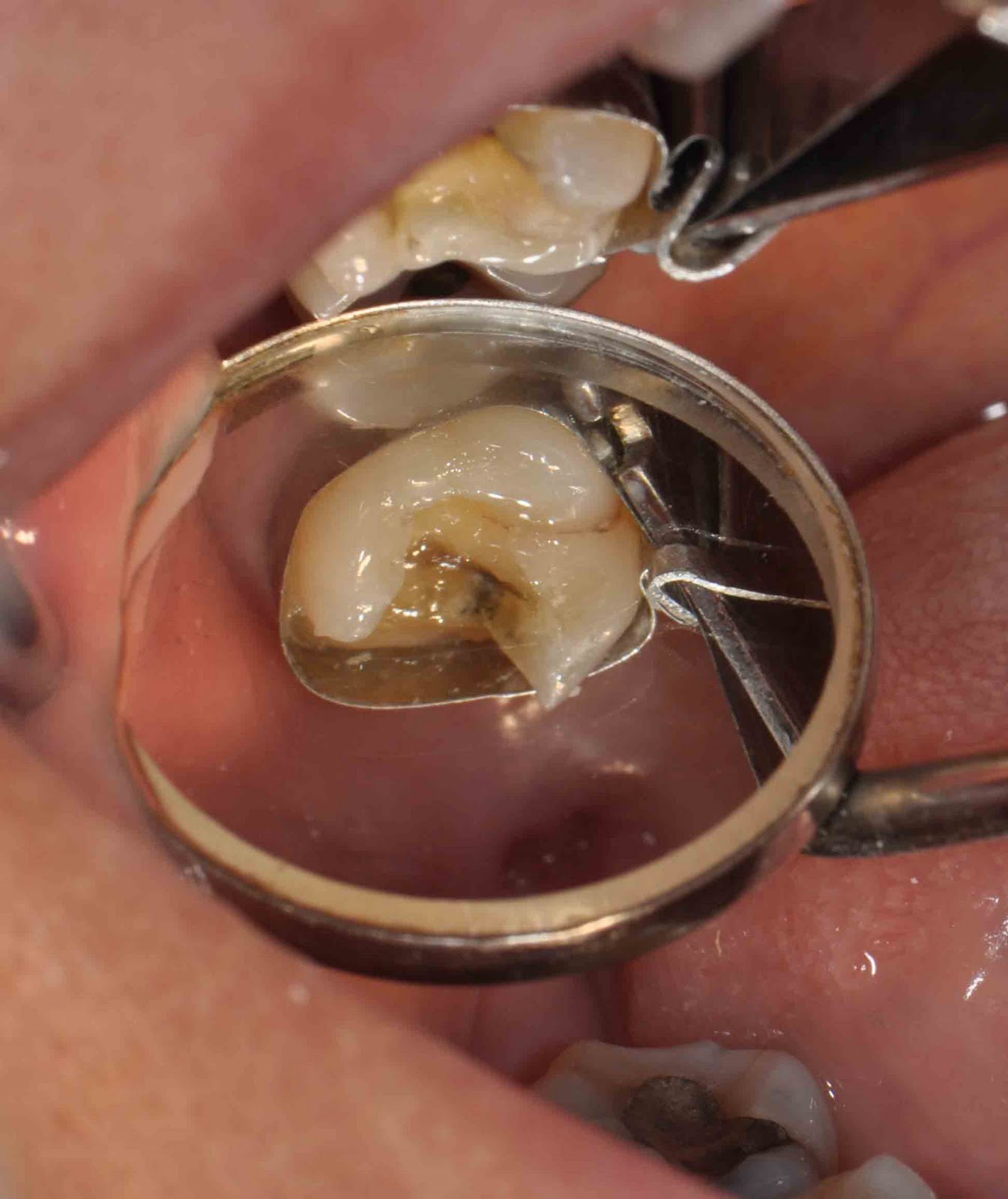
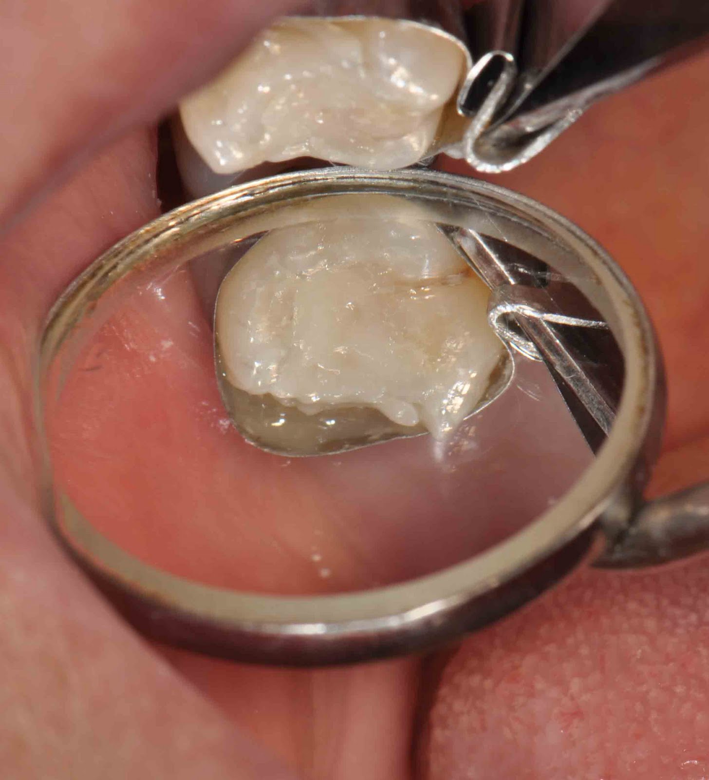
A) Placing the rubber dam.

After local anaesthetic is given, dentist usually need to “isolate” your tooth. He will first punch a small hole in a sheet of rubber. Then, he will then slip this sheet over the affected tooth and position a small tooth clamp to hold it there. The purpose of a rubber dam is to keep the tooth saliva-free therefore, avoid contamination of bacteria from saliva .

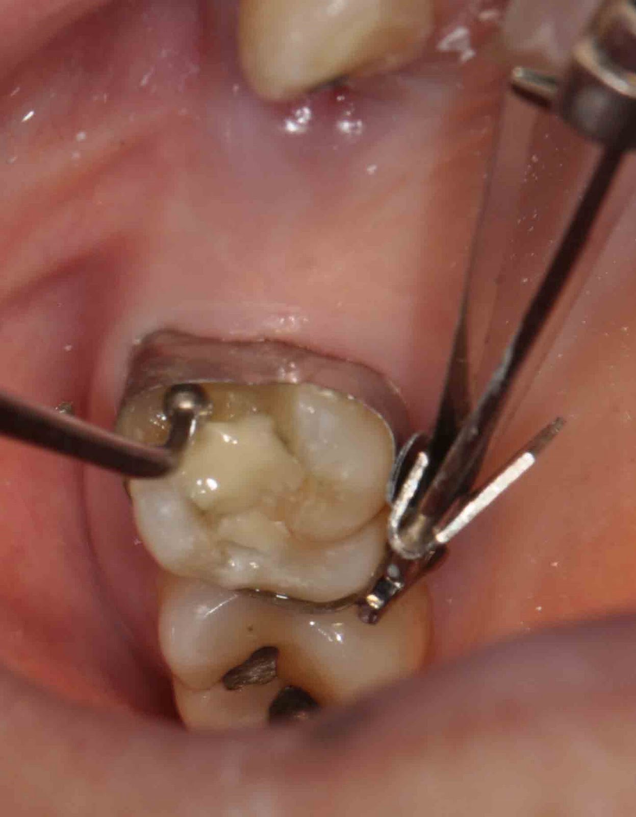
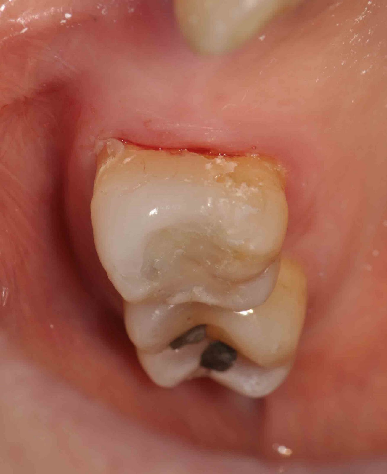
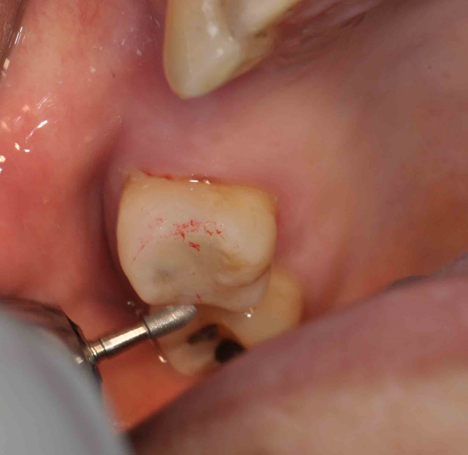
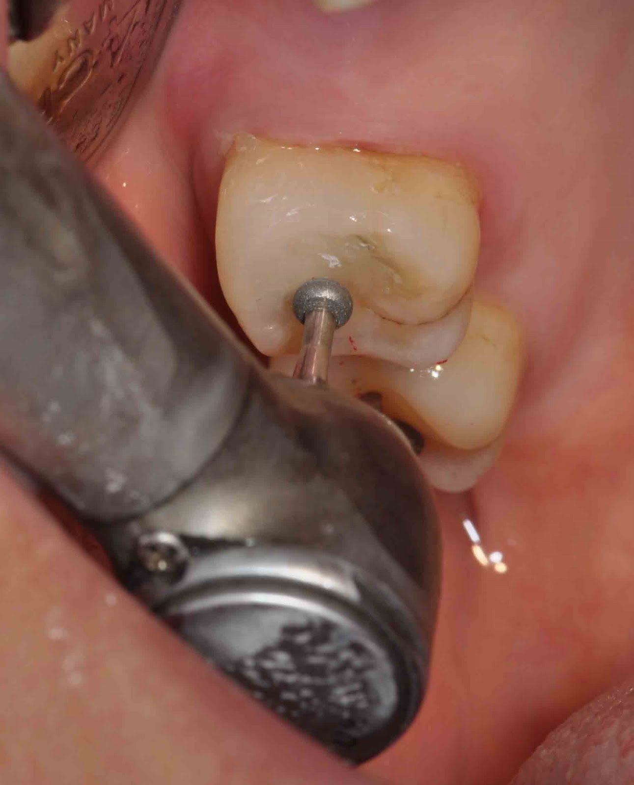
B) Creating the access cavity.

The tooth was isolated using rubber dam and a hole was made (cavity access) to reach the pulp chamber

As a starting point for performing  root canal treatment, dentist must first gain access to the nerve space (or the pulp chamber) within the tooth.
He do this by using a dental drill to create an access cavity. This hole will extend into the interior of the tooth to its pulp chamber. It’s the hole through which the dentist will perform their work.
On the molar teeth, the access cavity is made on the chewing surface of the tooth and for the front teeth, the access hole is made on the tooth’s backside.
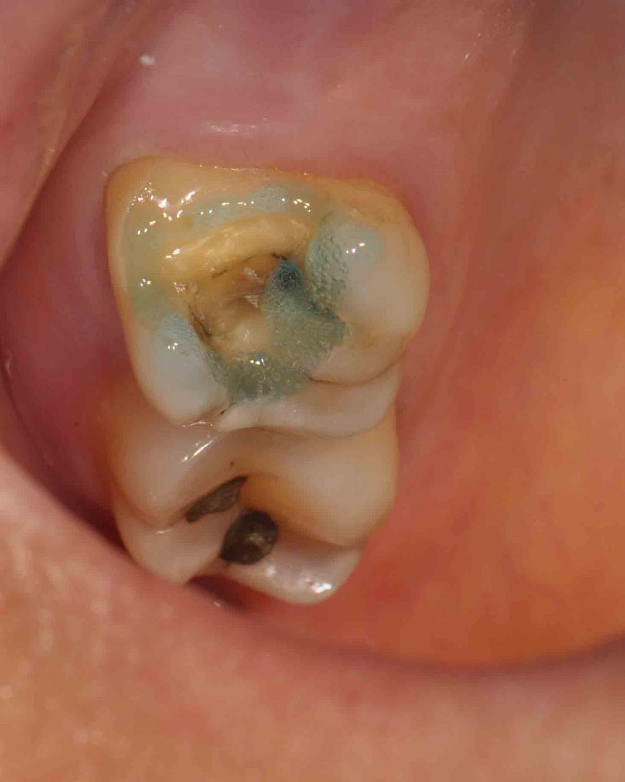
The overall size of the access cavity will vary according to factors such as the location of the individual canals and how hard it was for the dentist to find them. Additionally, beyond just that portion of the tooth that must be removed for access, the dentist will also need to remove any decay that’s present and any loose or exceptionally fragile tooth parts or fillings.

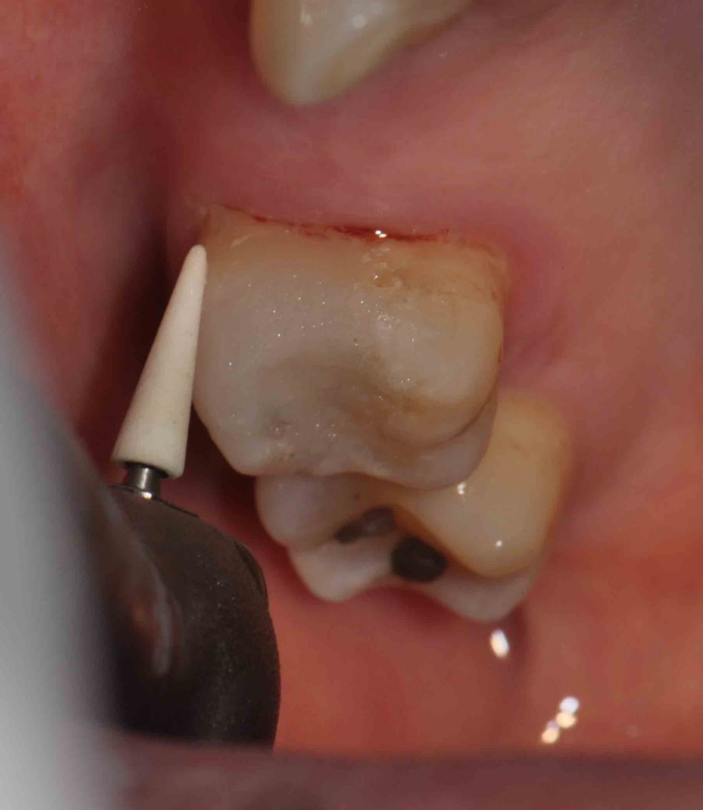
C) Cleaning and shaping the tooth’s root canals.

The next step of the root canal process involves “cleaning and shaping” the interior of your tooth (the pulp chamber and all root canals). In regard to the cleaning process, its purpose is to remove bacteria, toxins, nerve tissue, and related debris that are harbored inside the tooth.
The shaping process refers to how the tooth’s canals are enlarged and flared, so the have a shape that facilitates the filling and sealing process.

What instruments are used?

Endodontic file

For the most part, a tooth is cleaned and shaped using endodontic files. These files look like straight pins but on closer inspection you will find that their surface is rough, not smooth. These instruments literally are files and are used as such.

How are the files used?

Dentist works the file up and down, with a twisting motion, in each of your tooth’s root canals. This action will scrub, scrape and shave the sides of the canals, thus cleaning and sculpting them. He will perform this same type of action using a series of files, each having a slightly larger diameter.
The idea is that each consecutive file is used to slightly increase the overall dimensions of the root canal. Since some canal contaminates are embedded within a canal’s walls, this enlargement assists with both the procedure’s cleaning and shaping goals.
While performing this work, the dentist will also periodically flush out (irrigate) the tooth. This helps to wash away accumulated debris and contaminants. While a number of different solutions can be used for this purpose, sodium hypochlorite (bleaching agent) is the most common one. An added benefit of bleach is that it is a disinfectant.

Some dentist may have a handpiece that can manipulate the files for them.

Traditionally, files have been hand instruments. This simply refers to the fact that the dentist creates their filing action by manipulating them with their fingers. Some dentist may, however, have a special dental drill (handpiece) that produces the needed file motion for them.

Rotary Endodontic System

As a variation on this same theme, there is yet another type of dental handpiece that produces a cleaning motion by way of holding a root canal file and vibrating it vigorously.

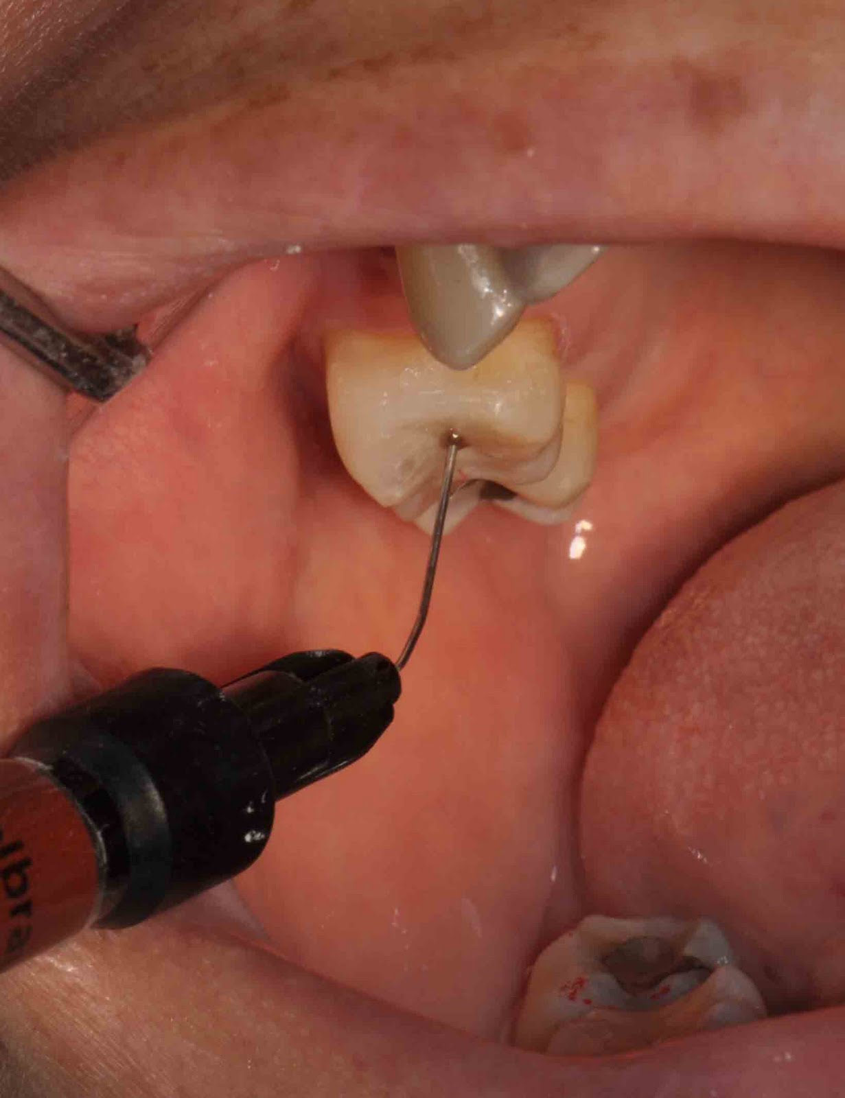
Measuring the length of the root canals.

Determine the length of root canal with a radiograph

The goal of root canal treatment is to achieve cleaning of the entire length of each of the tooth’s root canals, but not beyond.
As a means of determining the precise length of a canal, dentist will use apex locator to get the measurement for the length of the tooth (from the crown to the tip of the root). By doing so, the dentist wouldn’t go beyond during cleaning.
Usually, he will confirm the measurement by taking a x-ray of the tooth with a file placed in the tooth. The x-ray picture will show if the file extends the full length of the canal or not.

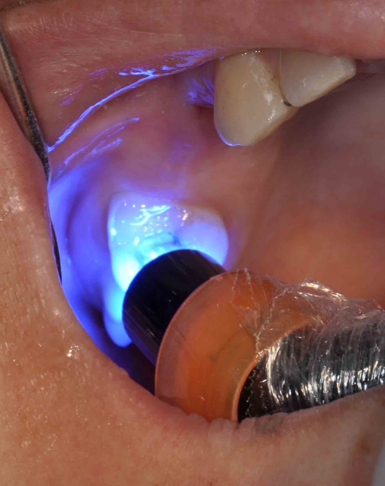
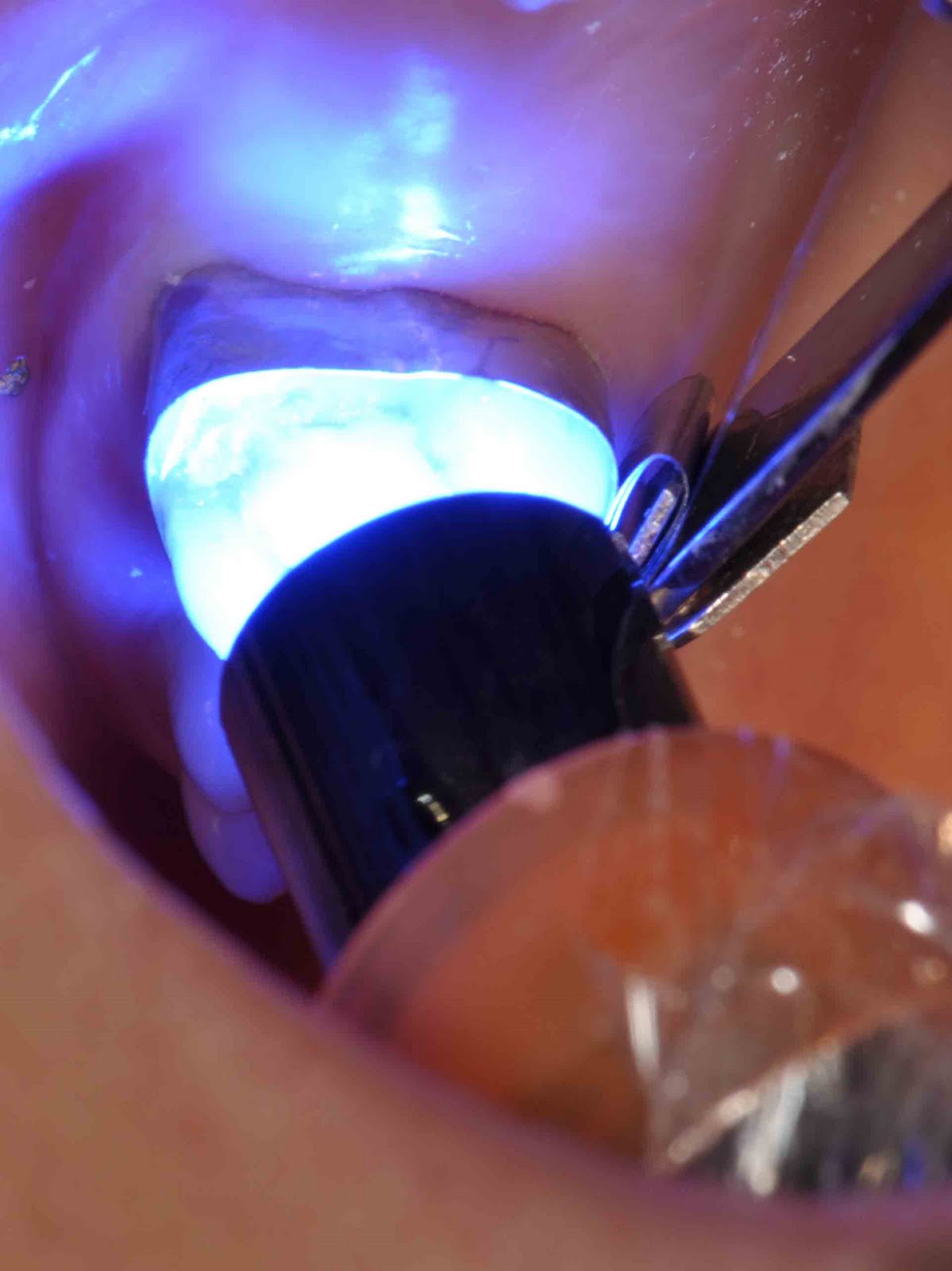
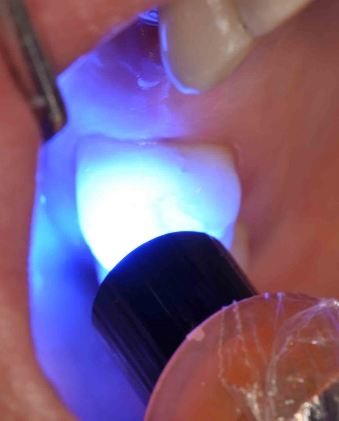
D) Sealing the tooth – Placing the filling material.

Once the interior of the tooth has been thoroughly cleansed and properly shaped, it is ready to be sealed (have its hollow interior filled in). In some cases, the dentist will want to place the filling material immediately after they have finished cleaning the tooth. With other cases, they may feel that it is best to wait about a week before performing this procedure.

What type of root canal filling material is used?

Sealing of the root canals

The most frequently used root canal filling material is a rubber compound called gutta percha. It comes in preformed cones whose dimensions match the size (diameter, taper) of the files that have been used to shape the tooth’s canals.
A root canal sealer (a paste) is usually used with the gutta percha. It is either applied to a cone’s surface before it is placed into a canal, or else applied inside the root canal itself before the cone is inserted. Several individual cones of gutta percha may be needed to fully fill the interior of the tooth.
Dentist will warm the gutta percha (either before or after it has been placed into the tooth) to soften it. This way it can be molded to closely adapt to the shape of the tooth’s interior.
As an alternative, a dentist may place the gutta percha via the use of a “gun.” This apparatus is somewhat similar to a hot-glue gun. It warms a tube of gutta percha. The softened material can then be squeezed out into the tooth.
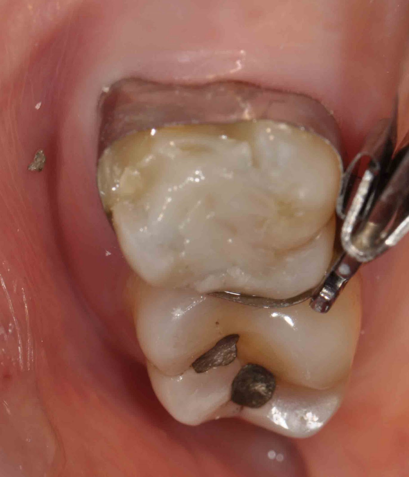
Once your dentist has finished sealing your tooth, they will place a temporary filling, so to seal off the access cavity created at the beginning of your treatment.

How long does root canal treatment take?

The total amount of time that’s needed for a tooth’s root canal therapy will of course hinge on how many appointments are needed (one visit, or two or more) and how long each one will take. Usually, root canal treatment for molar usually take about 3 to 4 visits. Each visit takes around an hour. For the front teeth, usually take fewer visits as they are simpler and located at the front region.

Summary of Root Canal Treatment:

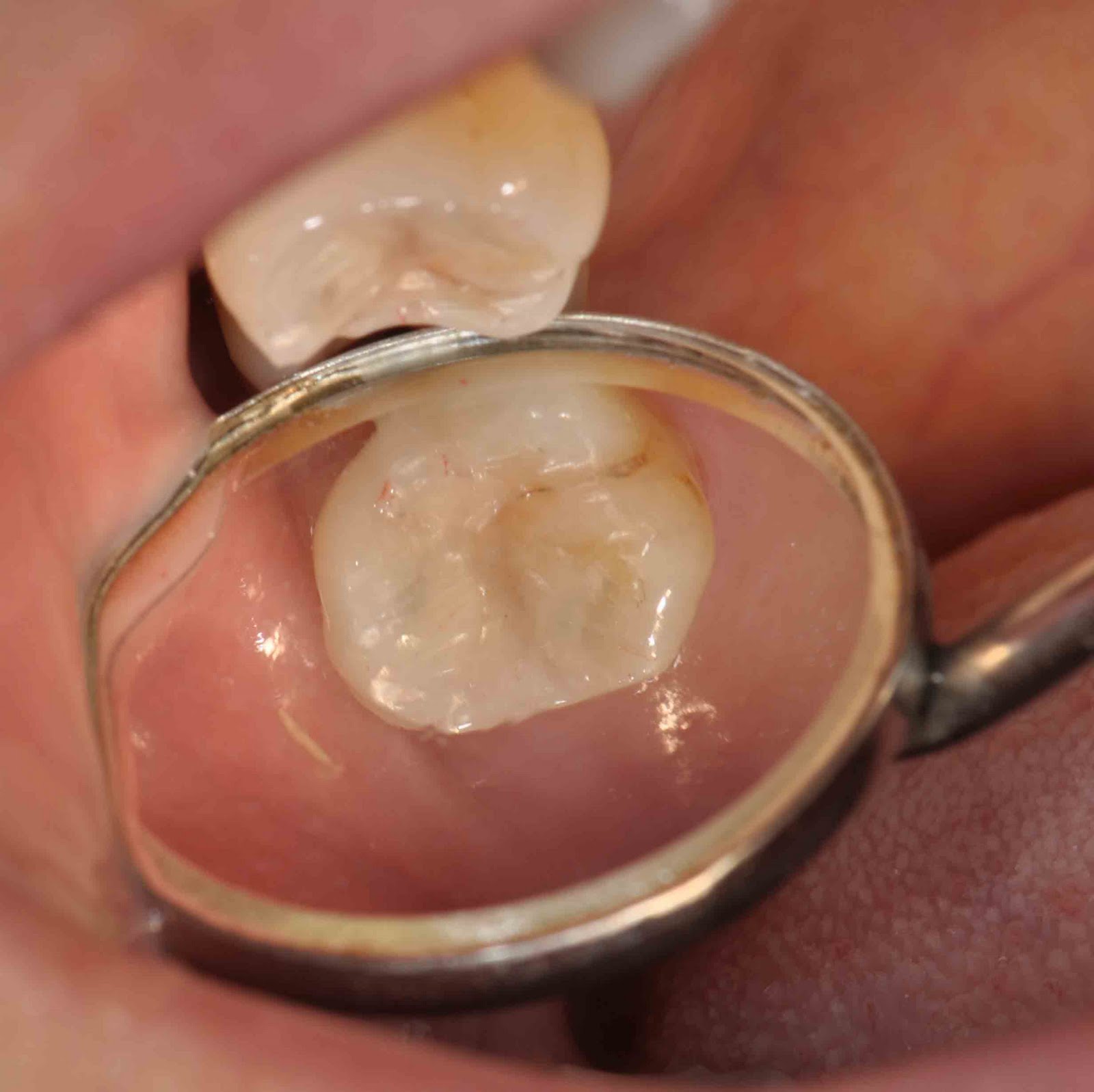
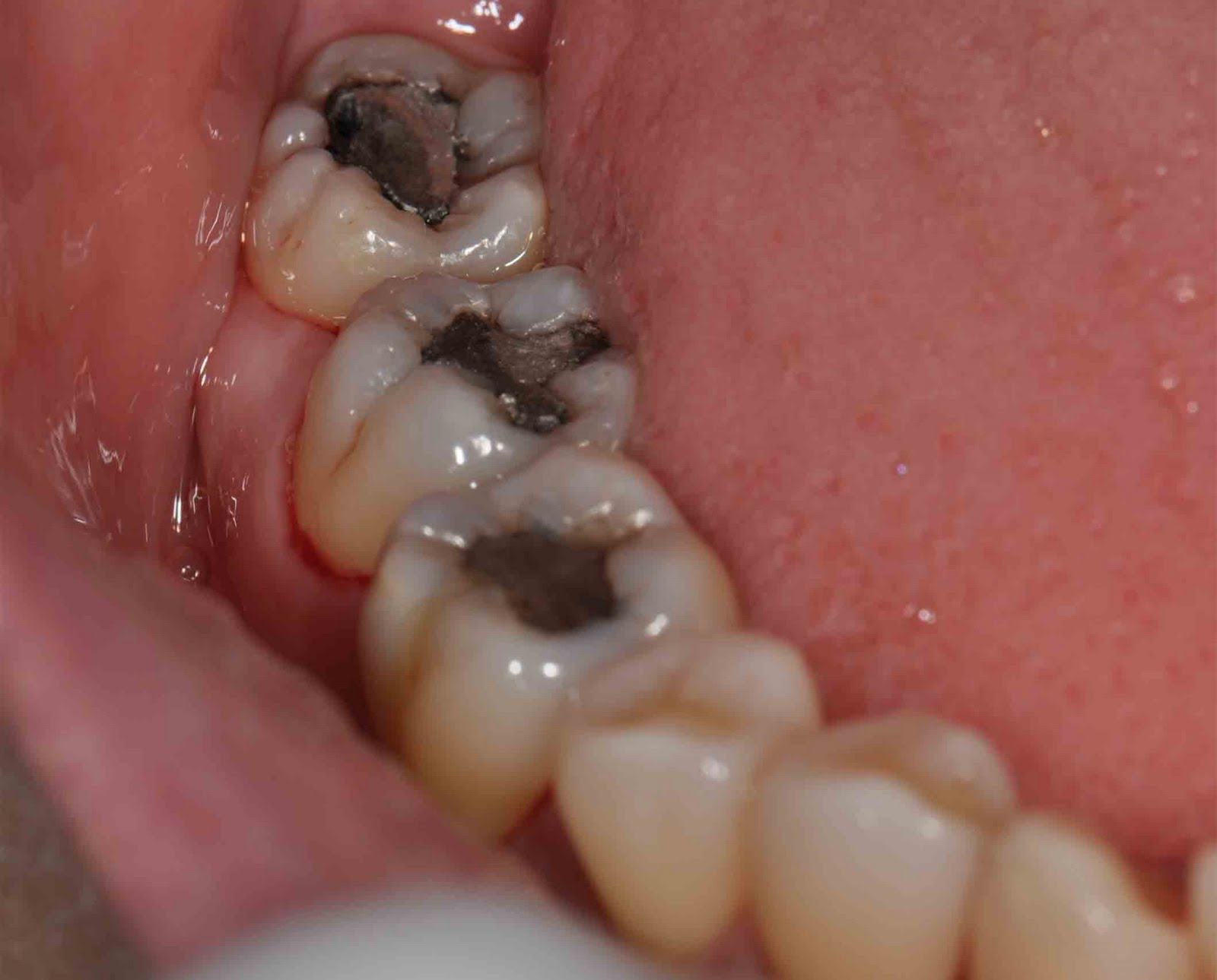
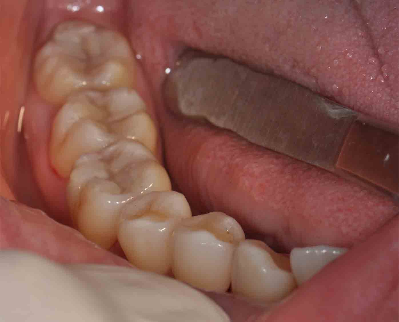
Cases of root canal treatments

RCT on upper left first molar
RCT on lower right first molar
RCT on lower rigth first molar


E) Post Root Canal Treatment

At this point, while the individual steps of performing the root canal process have been finished, the tooth’s treatment is not yet complete. The tooth is no longer alive since the pulp has been removed. And this can make the tooth brittle and more prone to fracture.
Therefore, a permanent restoration must still be placed. Choosing the right type of dental restoration, and having it placed promptly, will help to insure the long-term success of the tooth’s endodontic therapy.
There are a few options:
1) Crown (recommended) as it is strong, durable and more aesthetic
2) Dental filling – cheap, can be done on the spot (doesn’t required 2 visits)

F) Final words…

Tooth infection can recur in treated teeth (even RCT treated tooth), hence, good oral hygiene, including brushing, flossing and regular dental examination are necessary to prevent further problems. For more info on good oral hygiene click here.

More info on General Dental Treatment


20/Feb/2011

Periodontal-Health-&-Treatm
Root planing is a non-surgical treatment that usually done together with deep dental scaling. It is done after the supragingival (or superficial) plaque and calculus were remove through gross scaling. The aim of this treatment is to removes the roughened cementum and surface dentin that is impregnated with calculus, microorganisms and their toxins leading to the creation of a clean smooth root surface. Usually this implies that some cementum and dentin are removed. Deep scaling and root planing are done under local anaesthesia and requires 25-45 minutes per quadrant. If the pockets are deeper, the procedure becomes more significantly more difficult and more time is needed.


20/Feb/2011

Why do we have to clean our teeth?

Tartar (calculus) are deposits that have built up on the teeth over time. Dental cleanings involve removing plaque (soft, sticky, bacteria infested film) and tartar (calculus) deposits that have built up on the teeth over time. Your teeth are continually bathed in saliva which contains calcium and other substances which help strengthen and protect the teeth. While this is a good thing, it also means that we tend to get a build-up of calcium deposits on the teeth. This chalky substance will eventually build up over time. Usually it is tooth coloured and can easily be mistaken as part of the teeth, but it also can vary from brown to black in colour.

If the calculus (or tartar, as dentists like to call it) is allowed to accumulate on the teeth it will unfortunately provide the right conditions for bacteria to thrive next to the gums. The purpose of the scaling and polishing is basically to leave the surfaces of the teeth clean and smooth so that bacteria are unable to stick to them and you have a better chance of keeping the teeth clean during your regular home care. Also it leaves your teeth feeling lovely and smooth and clean, which is nice when you run your tongue around them.

10 Reason why you need to have Dental Cleaning


How are dental cleanings done?

Dental surgeon or dental hygienist uses specialized instruments to gently remove these deposits without harming the teeth.

1) Ultrasonic instrument (Scaler)

Ultrasonic Scaling

Commonly used first is an ultrasonic instrument or scaler unit which uses tickling vibrations to knock larger pieces of tartar loose. It also sprays a cooling mist of water while it works to wash away debris and keep the area at a proper temperature. The device typically emits a humming or high pitched whistling sound. This may seem louder than it actually is because the sound may get amplified inside your head, just like when you put an electric toothbrush into your mouth.

The ultrasonic instrument tips are curved and rounded and are always kept in motion around the teeth. They are by no means sharp since their purpose is to knock tartar loose and not to cut into the teeth. It is best to inform the operator if the sensations are too strong or ticklish so that they can adjust the setting appropriately on the device or modify the pressure applied.

With larger deposits that have hardened on, it can take some time to remove these, just like trying to remove baked-on grime on a stove that has been left over a long time. So your cleaning may take longer than future cleanings. Imagine not cleaning a house for six months versus cleaning it every week. The six-month job is going to take longer than doing smaller weekly jobs.

2) Air polishing

After the dentist has done scaling using the ultrasonic scaler, he will proceed with polishing either using air polishing or polishing cup. Air polishing is an alternative more advance method  than the polishing cup and paste method. It requires a special ultrasonic unit (e.g. Air Flow from EMS) that allows use of this insert in the handpiece.

EMS Air polishing unit

Air polishing uses medical-grade sodium bicarbonate and water in a jet of compressed air to “sandblast” the surface of the enamel leaving your teeth smooth and clean.

Indications for air polishing

  • Heavily smoking stain on the teeth
  • Staining due to coffee or tea
  • To remove fine tartar that are still attached to the teeth surface after scaling
Tobacco and coffee stain

Video: Air polishing method with EMS Air Flow®

Advantages of using air polishing

  • There is no physical contact with the tooth, therefore thermal injury is of no concern.
  • It is ideally suited for teeth separated by wide diastemata and considerable in shape and size.
  • Particularly good for cats where teeth are so small that standard cups can create gingival damage.
  • They are very efficient at removing stains from teeth.

Is scaling & air polishing going to be painful?

Most people find that cleanings are painless, and find the sensations described above – tickling vibrations, the cooling mist of water, and the feeling of pressure during “scraping” – do not cause discomfort. A lot of people even report that they enjoy cleanings and the lovely smooth feel of their teeth afterwards! There may be odd zingy sensations, but many people don’t mind as they only last a nanosecond.

Be sure to let your dentist/hygienist know if you find things are getting too uncomfortable for your liking. They can recommend various options to make the cleaning more enjoyable.

Painful cleaning experiences can be caused by a number of things: a rough dentist or hygienist, exposed dentine (not dangerous, but can make cleanings unpleasant), or sore gum tissues.

In case you may have had painful cleaning experiences in the past, switching to a gentle hygienist/dentist and perhaps a spot of nitrous oxide can often make all the difference. You could also choose to be numbed. If you find the scaling a bit uncomfortable because the gum tissues (rather than the teeth themselves) are sensitive, topical numbing gels can be used.

More info


Treatments of gum disease:


12/Feb/2011

Have your teeth checked regularly..


A standard dental checkup should take place every six months, and any additional procedures such as fillings or root canals, for example, require you to make additional appointments at later stage. This is because many of these procedures take longer than the time allotted to a checkup. They may even required preparation time on the part of the dentist, or time between appointments for the dental laboratory to manufacture crowns and dentures, among other fittings. Consequently, if your dentist finds any problems during the checkup, be prepared to visit him again in the near future.
At the routine checkup, the procedure is fairly simple. Generally, when reporting to the dentist’s receptionist, you are required to update your personal details and then wait to be called into the consulting or surgery room.
Once in the dentist’s chair, you are hoisted up and backward into a reclining position, and a light is positioned above you to afford the dentist the best view of your entire mouth. This is when you will be ask to relax – and open your moth wide!
The dentist will take a careful look at different parts of your mouth. He usually starts with your teeth, looking for any visible signs of disease. Then, taking a probe, he examines the surfaces of your teeth, keeping an eye out for any discolouration, soft spots and cavities.

Next he looks at the gum area to see if there are any signs of infection or recession, or bad deposits of plaque or tartar. He also looks for any lesions that might indicate more serious disease such as cancer, although these are rare. Overall, the dentist is assessing the quality of the gum tissue. Then, finally, he checks your tongue and palate to ensure they are healthy.
All of these procedures are completely painless; good news for those who avoid the dentist for most of their lives out of fear!

Patient position while taking X-ray

The next step is X-rays that are really quite quick and easy, although they may cause a little discomfort. This is because the X-ray plates are placed inside your mouth, and you have to bite down on them to hold them in place while the X-ray is taken. From time to time they may feel uncomfortable, or may press on your gums or teeth. However, they take only a couple of seconds each, and are important for your dentist to be able to see cavities in your teeth, as well as the state of each tooth in its entirety, and the bone.

Dental X-ray

Once your dentist has checked the X-rays, his assessment of your oral health is complete. Should you have no problems requiring treatment, he simply cleans and polishes your teeth and says goodbye until six months later. If you do have any disorders such as caries or gum disease, you will still go through the cleaning and polishing. However, your dentist will also spend some time with you outlining the treatment plan and discussing your options.

Read More

More info on General Dental Treatment


12/Feb/2011

Topics

  • Introduction
  • Reasons for Extraction
  • Types of Extraction
  • Simple Extraction
  • What can I Expect After an Extraction?
  • Instructions after tooth extraction

Tooth extraction

Introduction
Generally, dentists do everything within their power to preserve your natural teeth. However, in cases of advance caries or periodontitis, a tooth may have to be extracted. Teeth may also be removed for the purposes of orthodontics when teeth are straightened using braces. For example, your front teeth may be skew because there is not enough room for them in your mouth. An orthodontist may, therefore, suggest extraction of certain teeth to make space for teeth in your jaw.

Reasons for Extraction

The most most common reason for extraction is tooth damage due to breakage or decay. There are additional reasons for tooth extraction:

  • Severe tooth decay or infection.
  • Extra teeth which are blocking other teeth from coming in.
  • Severe gum disease which may affect the supporting tissues and bone structures of teeth.
  • In preparation for orthodontic treatment (braces)
  • Teeth in the fracture line
  • Fractured teeth
  • Insufficient space for wisdom teeth (impacted third molars).
  • Receiving radiation to the head and neck may require extraction of teeth in the field of radiation.
  • Deliberate, medically unnecessary, extraction as a particularly dreadful form of physical torture.

Types of Extraction

Extractions are often categorized as “simple” or “surgical”.
1.) Simple extractions are performed on teeth that are visible in the mouth, usually under local anaesthetic, and require only the use of instruments to elevate and/or grasp the visible portion of the tooth. Typically the tooth is lifted using an elevator, and using dental forceps, rocked back and forth until the Periodontal ligament has been sufficiently broken and the supporting alveolar bone has been adequately widened to make the tooth loose enough to remove. Typically, when teeth are removed with forceps, slow, steady pressure is applied with controlled force.
2.) Surgical extractions involve the removal of teeth that cannot be easily accessed, either because they have broken under the gum line or because they have not erupted fully. Surgical extractions almost always require an incision. In a surgical extraction the doctor may elevate the soft tissues covering the tooth and bone and may also remove some of the overlying and/or surrounding jawbone tissue with a drill or osteotome. Frequently, the tooth may be split into multiple pieces to facilitate its removal. Surgical extractions are usually performed under a general anaesthetic.

Simple Extraction

To extract a tooth, your dentist first administers a local anaesthetic in the area. Next, he firmly places extraction forceps over the crown of the tooth. He manually loosens the tooth, and then removes it. This is the most basic method of tooth extraction. Alternatively, he places an elevator between the tooth and the tooth socket, and carefully levers the tooth out.
However, sometimes it is impossible to remove a tooth using these methods, and then surgical intervention is required. Under local or general anaesthetic, the gum is cut over the relevant area, and the bone exposed. A section of bone is then removed to expose the root of the tooth, which is removed. Finally, the gum is stitched back together again.

What can I Expect After an Extraction?

It is critical to keep the area clean and prevent infection immediately following the removal of a tooth. Your dentist will ask you to bite down gently on a piece of dry, sterile gauze, which you must keep in place for up to 30 to 45 minutes to limit bleeding while clotting takes place. For the next 24 hours, you shouldn’t smoke, rinse your mouth vigorously, or clean the teeth next to the extraction site.

Sterile gauze to be placed at the extracted socket to stop bleeding

A certain amount of pain and discomfort is to be expected following an extraction. In some cases, your dentist will recommend a pain killer or prescribe one for you. It may help to apply an ice pack to the face for 15 minutes at a time. You may also want to drink through a straw, limit strenuous activity, and avoid hot liquids. The day after the extraction, your dentist may suggest that you begin gently rinsing your mouth with warm salt water (do not swallow the water). Under normal circumstances, discomfort should lessen within three days to two weeks. If you have prolonged or severe pain, swelling, bleeding or fever, call your dentist at once.

Instructions after tooth extraction

These are the instruction usually given to reduce complication such as excessive bleeding or infection after extraction:

  1. Please don’t spit or rinse after extraction for today.
  2. Make sure to bite gauze which ha s been placed over the extracted socket.
  3. Please bite the gauze for 30 minutes to allow bleeding ceased.
  4. Please not to change gauze too often.
  5. Take pain killer given if painful.
  6. Avoid taking food or drink which is too hot.
  7. If socket still bleeds; not to be too worry. You can rinse gently with some cold water.
  8. You can brush your teeth but gently and avoid the extraction site.
  9. On the following day, rinse with salt water.

Read more….


22/Jan/2011

Prestige Dental Care

Topics

  • To read on dental bridge click here
  • Crown – What is it?
  • How do we do it?
  • Picture on cases done in our clinic
  • Maintenance

Crown – What is it?

A crown is a replacement of the outside casing of your teeth that is permanently fixed to the tooth below. In other words, the decayed or damaged area of your tooth is removed and replaced by synthetic material, such as porcelain, to create a new ‘tooth’ with permanent results.

A crown is a replacement of the outside casing of your tooth

Crowns are often used after a root canal treatment, because the removal of a nerve causes a tooth to discolour. However, there are other reasons your dentist might crown one or more of your teeth: to restore a damaged or fracture tooth, to protect a weakened tooth so that it does not fracture under stress or to reinforce a large filling where there is not enough tooth structure left below.
Crowns are also made out of various material: gold or non-precious alloy, porcelain or ceramics, acrylic or composite resin, or even a combination of porcelain on metal. Obviously the material chosen will be in line with what will work best for your particular problem.

Gold crown

Non-precious Metal Crown

Porcelain fused with metal crown (PFM)

Full porcelain crown

Currently, most dentist use porcelain fused to metal crown or full porcelain crown as both crowns produce good aesthetic result

How do we do it?

When crowning a tooth, your dentist starts by filing away away between 1mm and 2mm on every surface of your teeth, and then the entire surface is smoothed. Next, an impression is taken of the prepared tooth by syringing an impression material into the space around the tooth and holding an impression tray in position until the material has set. This gives the laboratory a perfect negative copy of your tooth, which allows them to make up the permanent crown. The dentist also ensures he sends the laboratory details of what the colour porcelain to use, to ensure the crown looks natural.

1) Tooth are prepared and smoothen

2) Impression material is syringed into the space around the tooth to get the copy of the tooth

3) Negative copy of the teeth produced by impression material which is hardened on the impression tray

4) The crowns are fabricated on the model by laboratory technician

5) Finally, the crown is cemented onto the prepared tooth

Your are then fitted with a temporary crown, which is really just a rough space-filler, while you wait for the laboratory manufacture the permanent fixture.
At the following appointment, the dentist will have taken delivery of the permanent crown. The temporary crown is then removed, and the prepared tooth below thoroughly cleaned and dried. Then the new, permanent crown is cemented onto the tooth below. Any excess cement is removed, and the treatment completed.

Picture on cases done in our clinic

Here is a example of a discolour tooth that is replaced with a full porcelain crown (E-Max)

The young gentleman dislike his front discolour right central incisor

His tooth discolouration was due to old filling and the tooth is non-vital

After the tooth is prepared, colour of the porcelain crown is chosen to match his neighbour teeth

The full porcelain crown is cemented onto the tooth

Final Result – natural looking crown!!  (Treatment done by Dr. H.W.NG)

Maintenance

You do need to take care of your crown and not overstress it by biting or chewing very hard food. You can also floss normally around a crown. If at a later stage, your crown should break, you need to see your dentist as soon as possible, especially if the tooth inside becomes sensitive. Save the fractured piece if you can, as this can be glued back on temporarily until a new crown is made.

Read more

More info on General Dental Treatment


22/Jan/2011

Having to deal with a dental emergency is not something people think about. However, being prepared can make the difference between saving or losing a tooth. And in the case of a toothache, if it involves a bacterial infection, it can be a life-threatening situation.
Here are a couple of common dental emergencies and what to do about them.

A lost filling

Rinse out the cavity with warm water. Apply a temporary filling product such as Dentemp® which can be made into a ball and pressed firmly into the cavity. This can alleviate immediate pain, but it is important that you call your emergency dentist and arrange to have your filling replaced before the situation worsens.

Temporary filling

Lost Crown / Cap

Crown

Even though a missing crown is not immediately painful, it can become painful in a short period of time as food and other debris gather in the cavity. Unless the cavity is sealed up again, decay and infection can cause the tooth to ache. It is important that you visit your emergency dentist within a few days of losing your crown or cap.

You’re suffering with a toothache


Toothaches can be more dangerous than any physical trauma to a tooth. Rinse your mouth out with warm water and place some kind of cold compress against your cheek for twenty minutes to reduce the swelling. Then let it warm up for 20 minutes and then back to the cold compress.
Don’t put any kind of pain medication against the gum. The pain medication could burn the gum and cause more problems. The greatest risk comes if the toothache is from a bacterial infection. If left untreated, this could become life threatening. See an emergency dentist ASAP.
Source:
Shamblott Family Dentistry


11/Jan/2011

Topics
  • Introductions
  • Composition of composite resin
  • Restoring Composite Resin

Introductions

White filling or composite resin is getting more and more popular currently as it produces a nice aesthetic result. Previously silver filling (amalgam) is preferred due to low cost, easy to handle and it has a good mechanical properties (amalgam is strong and durable). However, current composite resin is as strong and durable as amalgam and on top of that, it produce a ‘hard to see restoration’ on the tooth making it a preferred choice for dentist and patient. Anyway, Nobody wants their filling to be seen!!

Composition of composite resin

Composite resins are composed of:
  • Bis-GMA monomers or some Bis-GMA analog
  • a filler material such as silica and in most current applications,
  • a photoinitiator
  • Dimethacrylates are also commonly added to achieve certain physical properties such as flowability.
  • Further tailoring of physical properties is achieved by formulating unique concentrations of each constituent.Unlike Amalgam which essentially just fills a hole, composite cavity restorations when used with dentin and enamel bonding techniques restore the tooth back to near its original physical integrity.
Variety of colours of composite resins
Empress Direct – Highly aesthetic composite material

Dental composite resin

In our clinic, we have multiple shade (or colour) to choose to mimic the original colour of the tooth in the mouth. For every restoration, we use different shade for each ‘layer’ during restoration.
Below are some of the cases done in our clinic.

Restoring Composite Resin

(Illustration in great depth)
An old amalgam was removed from a upper right molar and the tooth was ready for filling placement
Firstly, the enamel surface of the molar was treated with phosphoric acid 35%
Then, it was followed by the dentine which was located at the center of the tooth
Next, the cavity was cleaned and dried. A thin layer of adhensive solution was applied over the cavity
Light cure unit was used to activated the adhesive which was photo-sensitive
A metal band was used to wrapped around the molar
The metal band was checked to ensure that there was no gap between the tooth and the metal band. This was to prevent the filling material from overflow out from the cavity.
‘Dentine shade’ composite resin was placed at the center of the cavity to cover the darkish stain in the dentine area.
The composite was light-cured to make it harden
Next, ‘enamel shade’ composite was placed over the ‘dentine shade’ composite (layering technique) to get the translucency effect.
‘Enamel shade’ composite was placed at the side-wall of the molar and was cured (with light-cure unit)
The metal band was removed and more composite was placed at the side-wall of the molar so to achieve a nice contour and bulbosity.
This was the result after the composite harden
The composite excess was removed with a rotary white stone
The restoration was checked to look of  any interference with the lower teeth during biting and chewing (with a bite registration tape)
And the red markings from bite registration tape was removed
Finally, for aesthetic reason, grooves and fissures was make on the tooth surface
And ‘stain’ composite was placed on the fissure to make it looked like the neighboor teeth
Lastly, light-cure unit used to harden the rest of the composite
The restoration was polished to make is glossy and shining
The Final Result!!
Before and after picture.

Another case: Replacing old silver (amalgam) restorations with composite restorations

This young gentleman came complaining of sensitive to hot and cold at his bottom right molars. On examination, he had a few siler fillings which cracked and had gaps noted at the margin fo the fillings.
The silver fillings were removed and replaced with white fillings. The fillings match with the colour of the tooth  very well as if there was no filling done!!

03/Jan/2011

Topics

  • What is Tooth Decay?
  • How Will I Know if I Have a Cavity?
  • How Do Dentists Detect Cavities?
  • Are Some People at More Risk for Developing Cavities?
  • How Can I Prevent Cavities?
  • What should I do if I have tooth decay?

What is Tooth Decay?

Dental cavities are an infection caused by a combination of carbohydrate-containing foods and bacteria that live in our mouths. The bacteria are contained in a film that continuously forms on and around our teeth. We call this film plaque. Although there are many different types of bacteria in our mouths, only a few are associated with cavities. Some of the most common include Streptococcus mutans, Lactobacillus casei and acidophilus, and Actinomyces naeslundii.

When these bacteria find carbohydrates, they eat them and produce acid. The exposure to acid causes the pH on the tooth surface to drop. Before eating, the pH in the mouth is about 6.2 to 7.0, slightly more acidic than water. As “sugary foods” and other carbohydrates are eaten, the pH drops. At a pH of 5.2 to 5.5 or below, the acid begins to dissolve the hard enamel that forms the outer coating of our teeth.
As the cavity progresses, it invades the softer dentin directly beneath the enamel, and encroaches on the nerve and blood supply of the tooth contained within the pulp.
Cavities attack the teeth in three ways:
1. Pit & Fissure
2. Smooth surface
3. Root surface

The first is through the pits and fissures, which are grooves that are visible on the top biting surfaces of the back teeth (molars and premolars). The pits and fissures are thin areas of enamel that contain recesses that can trap food and plaque to form a cavity. The cavity starts from a small point of attack, and spreads widely to invade the underlying dentin.

Decays formed at the the pits and fissures and spread to the whole biting surface of the teeth

The second route of acid attack is from a smooth surface, which is between, or on the front or back of teeth. In a smooth-surface cavity, the acid must travel through the entire thickness of the enamel. The area of attack is generally wide, and comes to a point or converges as it enters the deeper layers of the tooth.

At the smooth surface area, decay started in-between of the teeth

The third is the attack started at the root surface of the tooth after it was exposed to the oral cavity. The root is usually exposed due gum recession as a result of periodontitis (gum disease)

Decay at the root surface of the teeth

Recipe for tooth decay formation?

How Will I Know if I Have a Cavity?

The large majority of cavities are completely painless. This is because the outer enamel has no nerves. It is only when the cavity enters the underlying dentin that the cavity may begin to feel sensitive (Teeth sensitive). The most common cavity symptoms are an increased sensation to cold, sweet foods or beverages. A cavity is often responsible for a broken tooth. The cavity weakens the tooth, especially when it forms under a tooth filling or a tooth cusp, and can easily cause a fracture when biting down.

Sensitive Teeth

Patients are sometimes taken off guard when they learn that they have a few cavities but they don’t have any symptoms. It is far better to treat a small cavity than to wait until they have symptoms; such as pain. By the time there are symptoms, the cavity may have spread to infect the dental pulp, necessitating a root canal procedure or a tooth extraction to eliminate the infection. Always remember that most dental problems are insidious — that is, they sneak up on you. Regular dental exams, at least twice a year, will greatly reduce the likelihood that a dental cavity will go undetected and spread, causing toothache pain and infecting the dental pulp.

The decay has spread into dental pulp causing pain

How Do Dentists Detect Cavities?

Cavities are detected a number of ways. The most common are clinical (hands-on) and radiographic (X-ray) examinations. During a clinical exam, the dentist uses a handheld instrument called an explorer to probe the tooth surface for cavities. If the explorer “catches,” it means the instrument has found a weak, acid damaged part of the tooth — a dental cavity. Dentists can also use a visual examination to detect cavities. Teeth that are discolored (usually brown or black), can sometimes indicate a dental cavity.

Regular dental examination is important to prevent tooth decay

Bite-wing radiograph is good to detect interproximal (in-between) caries

Dental X-rays, especially check-up or bitewing X-rays, are very useful in finding cavities that are wedged between teeth, or under the gum line. These “hidden” cavities are difficult or impossible to detect visually or with the explorer. In some cases, none of these methods are adequate, and a dentist must use a special disclosing solution to diagnose a suspicious area on a tooth.

Are Some People at More Risk for Developing Cavities?

People who have reduced saliva flow due to diseases such a Sjogren Syndrome; dysfunction of their salivary glands; have undergone chemotherapy or radiation; and who smoke are more likely to develop cavities. Saliva is important in fighting cavities because it can rinse away plaque and food debris, and help neutralize acid. People who have limited manual dexterity and have difficulty removing plaque from their teeth may also have a higher risk of forming cavities. Some people have naturally lower oral pH, which makes them more likely to have cavities.

How Can I Prevent Cavities?

The easiest way to prevent cavities is by brushing your teeth and removing plaque at least three times a day, especially after eating and before bed. Flossing at least once a day is important to remove plaque between your teeth. You should brush with a soft-bristled toothbrush, and angle the bristles about 45 degrees toward the gum line. Brush for about the length of one song on the radio (three minutes). It’s a good idea to ask your dentist or hygienist to help you with proper brushing methods.

Blushing and Flossing teeth are to do it daily to stop caries

Reducing the amount and frequency of eating sugary foods can reduce the risk of forming cavities. If you are going to drink a can of sweetened soda, for instance, it is better to drink it in one sitting, than sip it throughout the day. Better yet, drink it through a straw in one sitting, to bypass the teeth altogether. Getting to the dentist at least twice a year is critical for examinations and professional dental cleanings.

Reduce high sugar food can reduce dental cavity significantly
To reduce the incidence of cavities, use toothpaste and mouthwash that contains fluoride. Fluoride is a compound that is added to most tap water supplies, toothpastes, and mouth rinses to reduce cavities. Fluoride becomes incorporated into our teeth as they develop and makes them more resistant to decay. After our teeth are formed, fluoride can reverse the progress of early cavities, and sometimes prevent the need for corrective dental treatments.

Mouthwash with fluoride

The recent drop in the number of cavities is largely due to the addition of fluoride to our drinking water. Mass water fluoridation is the most cost-effective measure available to reduce the incidence of tooth decay. The Environmental Protection Agency has determined that the acceptable tap water concentration for fluoride is 0.7 to 1.2 parts per million.

A dental procedure called sealants can also help reduce cavities on the top and sides of back teeth (occlusal, buccal and lingual surfaces). A sealant is a white resin material that blankets the tooth, protecting the vulnerable pits and fissures of the tooth. Sealants are routinely placed on children’s teeth to prevent cavities on their newly developing molars. The use of sealants to prevent cavities is also a cost-effective way to reduce the incidence of cavities on adults as well. Sealants are generally not used on teeth that already have fillings.

Fissure Sealant

People who have a dry mouth are at risk for developing cavities, and can have their dentist prescribe artificial saliva and mouth moisturizers, as well as recommend chewing sugarless gum to stimulate saliva production. Finally, an antiseptic mouthwash containing chlorhexidine gluconate such as Chlohexxa or Oradex can also be useful in killing bacteria associated with dental caries.

What should I do if I have tooth decay?

You should go the to dental clinic as soon as possible. Early or small decay is easily to treat. Usually a small filling will do. However if it is large cavity, then a larger filling is required provided there is no pain. In cases where the tooth is painful (eg. pain on biting, disturb sleep), then root canal treatment or extraction is required to stop the infection.
Small filling
Filling can be silver (amalgam) or white (composite).
Large Filling
Usually required
Comparison within big and small filling:
Small Filling                           vs.       Large filling

  • Less pain during filling                More pain (because lager & deeper cavity)
  • More aesthetic                             Less aesthetic
  • More lasting and durable             Less durable
  • Cheaper                                       More expensive (more filling material)

Crown
Or tooth capping of is a procedure to created back function, aesthetic as well as protection to a severely damaged tooth. It is usually made of porcelain fused with metal or a full porcelain material. Crown is durable and more lasting compared to a large filling.

Crown

Root canal treatment (RCT)
RCT is required when infection from caries has spread to the pulp of a tooth. The tooth is usually painful on chewing and sometimes disturb sleep. The purpose of this treatment  is to preserve the tooth by removing the dead and infected pulp leaving the tooth bacteria free.
After RCT, the tooth can be restored with filling or a corwn. If there is a lot of tooth structure loss, the tooth should be protected with a crown.

Root Canal Treatment

Extraction
Tooth extraction in another way to stop infection. However, this method is commenced  if patient don’t want to keep the tooth anymore. Patient have to understand the consequent of removing the tooth
Root canal treatment              vs.        Tooth extraction

  • Tooth preserved                             Tooth removed
  • Difficult (esp molar tooth)              Simple & fast
  • Expensive                                       Cheaper than RCT
  • Few visits                                       One visit
  • Lesser problems in future              More problems in future

Tooth Extraction

Read More


02/Jan/2011

Topics

  • What is dental fillings?
  • The goal of treating cavities
  • What Dental Material is Best for My Teeth?

What is dental fillings?

Dental filling is a dental restorative material used to restore the function, integrity and morphology of missing tooth structure. Basically… material used to fill up cavity in the tooth after the decay was removed from the tooth.

Most cavities discovered during a dental examination will need to be treated. In general, if a cavity has broken through the enamel and is into the underlying dentin, or is able to be probed with an explorer, it has undergone cavitation and requires treatment. Early dental cavities that have not spread to the dentin or have undergone cavitation should not be treated, as they can be healed or re-mineralized with fluoride.

The goal of treating cavities involves two basic principals:

1. Removing the decayed portion of the tooth
Teeth Decay
After the decay portion is removed and the tooth is ready for filling
2. Rebuilding the missing tooth structure with a filling material.
Filling material is used to replace missing tooth structure due to decay
The dentist usually begins the procedure with an injection of local anesthetic if the decay is deep and very sensitive. A high-speed dental drill is needed to remove the decay and prepare the tooth for the filling. Depending on which material is used, the dentist will vary the tooth preparation accordingly.
Dental drill (High-speed handpiece)
Decay is removed with a high-speed dental drill
After the tooth has been prepared, a liner (Dycal®)is often used to reduce tooth sensitivity.Dycal is a compound containing calcium hydroxide, and is used in deep cavities to stimulate the dentin to regenerate and protect the dental pulp. In deeper fillings, a base is used in addition to the liner. Common bases used under dental fillings are glass ionomer cement and zinc phosphate cement. The main purpose of the base is to insulate the tooth from temperature changes in the mouth. The dentist and patient can then choose a number of different materials to fill the tooth, but the most common are silver (amalgam), white (resin), porcelain or gold. These materials are layered on top of the liner or base to finish the process of rebuilding the tooth. After a tooth has been filled, it is not unusual for the tooth to be sensitive for a day or two. In general, the deeper the filling, the more likely the tooth will have prolonged sensitivity, especially to cold food or beverages. Most fillings should be completely comfortable within two weeks. In some cases, the filling will be built up too high, and a second appointment is needed to shave down the filling to a comfortable level. If sensitivity lasts more than two weeks, it may indicate that there is a void under the filling. Prolonged discomfort may also indicate a tooth that has an infected pulp, and requires root canal therapy.

What Dental Material is Best for My Teeth?

Dental Amalgam

Silver (Amalgam) Filling
Every dental material used to rebuild teeth has advantages and disadvantages. Dental amalgam or silver fillings have been around for over 150 years. Amalgam is composed of silver, tin, copper, mercury and zinc. Amalgam fillings are relatively inexpensive, durable and time-tested. On the flip side, they are considered unaesthetic because they blacken over time and can give teeth a gray appearance, and they do not strengthen the tooth. Some people worry about the potential for mercury in dental amalgam to leak out and cause a wide variety of ailments, but research does not bear this fear out. Advantages:
  • Cheap
  • Strong
  • Lasting
  • One visit
Disadvantages:
  • Not aesthetic (Silver colour)
  • Blacken over time and can give the teeth a gray appearance
  • Contain mercury
  • Required deeper cavity to retain amalgam
  • No chemical bonding to tooth structure

Composite Resin/White Filling

White (Composite Resin) Filling
Composite resin, or white fillings have been around for about two decades. Composite fillings are composed of an organic polymer known as bisphenol-A-glycidyl methacrylate (BIS-GMA), and inorganic particles such as quartz, borosilicate glass and lithium aluminum silicate. They have the advantage of requiring a more conservative tooth preparation (less drilling required), can have a strengthening effect on the tooth and are very aesthetic, virtually blending in with the tooth. Composite fillings are the material of choice for repairing the front teeth. On the down side, they are more technique-sensitive for the dentist to place, and are highly susceptible to decay in the future if placed improperly. They usually cost more than an amalgam. Despite this research composite fillings are considered safe, and like the other dental filling materials, they are approved by the American Dental Association. Advantages:
  • Highly aesthetic – it can be used to improve aesthetic
  • Can be used for shallow cavities
  • Chemically bond to tooth structure
  • One visit
Disadvantages:
  • Technique sensitive (required dry surface for filling adhesion)
  • Can result in tooth sensitivity (due to shrinkage of the composite)
  • The strength of composite is lesser than amalgam
  • Required replacement or repair due to staining, chipping, wear and tear
  • Cost more than amalgam
More info on aesthetic filling click here

Porcelain inlay/onlay/crown

Porcelain onlay used to formed back missing tooth structure
Porcelain crown
Porcelain crown
Porcelain is sometimes used for dental fillings called onlays or inlays. Porcelain is a non-crystalline glass composed of silicon and oxygen. It has the advantage of being highly aesthetic, and is the restoration of choice for people who place the highest value in the appearance of their teeth. Porcelain has the disadvantage of being brittle, and, therefore, susceptible to breakage. It is also even more technique-sensitive to use than composite; requires two dental visits to place the filling; and costs significantly more than amalgam or composite fillings. Porcelain can also cause accelerated wear of the opposing tooth when biting. Advantages:
  • Highly aesthetic
  • Strong
  • Lasting
Disadvantages:
  • Required to scarified more tooth structure for retention
  • The retention of the porcelain is depends on the cement used to ‘glue’ the porcelain to the tooth
  • Porcelain is brittle and susceptible to breakage
  • Required two visits
  • Can cause accelerated wear of the opposing tooth when biting
  • Expensive
More info on crown click here

Gold Inlay

Gold onlay
Gold is sometimes used for dental fillings, most commonly as an inlay. Gold is not used in its pure form, but as an alloy containing 75 percent gold, as well as copper, silver, platinum, palladium and zinc. Gold is extremely durable; fairly aesthetic, it does not damage the opposing tooth when biting, and is very well tolerated by the gums and other intraoral tissues. A well-done gold filling can last two to four times longer than any other dental material and might be considered the “gold standard” for dental fillings. Gold inlays, like porcelain inlays, take two dental visits to complete and are also much more costly than amalgam or composite. They are also not nearly as aesthetic as composite or porcelain. In addition, gold inlays are fairly difficult to prepare and place.

Advantages:

  • Durable
  • Strong
  • Lasting
  • Very well tolerated by the gums and other intraoral tissues (bio compatibility)
  • Does not damage the opposing tooth when biting (as compared to porcelain)

Disadvantages:

  • Not aesthetic
  • Difficult to prepare
  • Required to scarified more tooth structure for retention
  • Expensive because it is GOLD

Read more

More info on General Dental Treatment


Get in touch

Contact us now

Book an appointment with our doctors now!! Click here
If you have any question, don’t hesitate to contact us, we are more than glad to provide you with the information you need!

We accept Cash, Credit Card, Grabpay, Alipay, Touch n Go, MayBank QRPAY and Boost


Follow us

Our Activity

Curious about what we do every day? Follow us to learn more about the dental health care we provide.


Copyright 2023. All rights reserved.