Blog

NEWS AND DENTAL EDUCATION

We share informative articles and news.


22/Jan/2021


Oral cysts and tumors may develop in the jawbone or soft tissues in the mouth and face.
Oral cyst is an abnormal cavity within bone or soft tissues which may contain fluid. There are many different types of cysts in the mouth.

Reason to remove oral cyst:

  • Size of cyst will increase with time
  • Very large cyst can be dangerous, they can weaken your jawbone and causes your jawbone to be break easily
  • Teeth adjacent to large cyst can be affected and become mobile

Oral tumor is a solid or semi-solid mass in the bone or soft tissue that is made of unusual cells. Oral tumor can be benign tumor (not cancerous) or malignant tumor (cancerous). This can be confirmed by using biopsy test (a surgery that removes part of your tumor to examine it under a microscope). Biopsy is needed to diagnose the tumor before starting any treatment.

When should I suspect that I have an oral cyst or tumor?
Most of the oral lesions do not have any symptoms. Oral cyst may cause swelling, bone expansion, displacement or loosening teeth and pain (if infected). Oral tumor may present as a non-painful bump, lump, or ulcer. After your oral and maxillofacial surgeon examines the cyst or tumor, he will often recommend an X-ray to determine what kind of treatment you need.

Type of common treatments for oral tumors and cysts – Treatment options depends on the location and the type of lesion and your symptoms.

  • Fine needle aspiration cytology (FNAC): FNAC is a fast and less invasive tool used to diagnose a suspicious lesion or to differentiate cancerous and non-cancerous lesion before any invasive surgery done. This technique uses fine needle to obtain cells from lesion for examination under a microscope or to explore whether a lesion contains a fluid.

  • Surgical excision: Complete removal of lesion with the surrounding normal tissues.
  • Incisional biopsy: This technique removes only a small portion of a large lesion for diagnosis prior to treatment. Incisional biopsy indicated in very large lesion, hazardous location of the lesion and lesion that has the characteristics of malignancy.

  • Enucleation: A technique used to completely remove cyst by elevating of soft tissue flap, removing of bone overlying the cyst followed by peeling off the cyst with spoon-like instrument.

  • Marsupialization: Removes only a portion of the cyst lining and the cystic lining is sutured to the oral mucosa to keep the cavity open. The open cavity will trap food and takes many months to heal. This technique rarely used, unless associated structures, such as adjacent nerve, nose, and maxillary sinus are at the risk of damage during enucleation.

The post was prepared by Dr. Jean

Read More…


22/Jan/2021

Multiple dental extraction is defined as removal of more than one tooth in a single dental visit. This procedue usually will be done under general anaesthesia (GA) in the hospital setup. Dental extraction under GA also can be performed on children who is anxious, uncooperative or having medical condition such as cerebral palsy or hyperactive child. In some cases, dental extraction under GA is done on children with badly decay teeth which needed multiple dental visit for extraction. It is therefore, better to have it done under general anaesthesia.

The indications of dental extractions under general anesthesia are:

    • Young child with rampant decayed teeth
    • Children who is unsuitable for local anesthetic or conscious sedation due to lack of cooperation
    • Patient with medical condition such as cerebral palsy or severe seizure
    • Patient who required lot of tooth extraction


Dental extraction under GA di performed in the hospital setup as day-care (no hospitalization). The patient (or the child) is usually health (ASA I) and fasting since midnight. The anesthesiologist will assess the child and written consent is taken before the procedure started.

In the operating room, the anesthesiologist will put a tube in patient’s throat to administer ‘sleeping’ gas allowing patient to ‘sleep’ during the whole procedure. The oral surgeon will come in to perform the dental extraction. After the procedure, the tube with be removed from the patient’s throat, and he will placed in the recovery room. He will be allowed to discharge from the hospital when fully conscious and ambulated.

Upon the procedure, a minor bleeding in extraction site, swollen tongue/lip/chin/cheek, temporary mouth opening limitation are the sequelae or conditions to be expected.

Advantage of extraction under GA

  • Very useful to traet uncooperative patient
  • Patient has no experience of the awlful dental extraction
  • No need many dental visit for extraction
  • Other dental procedures such as fillings and dental cleaning can sometimes be done in the same visit

Disadvantage of extraction under GA

  • Patient has to be healthy for GA
  • The procedure has to be done in the hospital setup with OT facility
  • Treatment will cost more as the patient has to be admitted in the hospital as well as GA fees

Read more….


02/Dec/2020

Minor oral surgery is a field in dentistry that involves surgical treatment that performed within the mouth. This surgery can be done under local anaesthesia with or without sedation. In most situations, it requires only a relatively short recovery period. Types of Minor Oral Surgery

  • Impacted wisdom teeth removal – Surgery to remove wisdom teeth
  • Difficult Teeth Extaction (Surgical Extraction) – extraction that require the elevation of soft tissue flap, removal of bone, and/ or sectioning of the tooth
  • Apicectomy – removal of the last portion of the root of a teeth
  • Cyst Enucleation – a technique used to completely remove cyst by elevating of soft tissue flap,  removing of bone overlying the cyst followed by peeling off the cyst with spoon-like instrument.
  • Gum Surgery
  • Dental Implant Treatment – placement of dental implant (that look and feel like your natural teeth) to replace your missing tooth.
  • Biopsy – is a procedure where a small part of tissue is removed so that it can be looked closely under microscope.


16/Jul/2013

Periodontal-Health-&-Treatm

After the age of 35, gum disease or periodontitis is the major cause of tooth loss in adults, far more so than tooth decay. In fact, about 80% of tooth loss can be ascribed to periodontal disease in this age group. A lot of time and money could be saved by early detection and treatment of the disease and many more people would keep their teeth if they were aware of this fact.

Sequela of gum disease

Periodontal disease affects the support structures of the teeth: the bone, gums and ligament (Click here for Dental Anatomy). It is long-term and slow-moving disease: painless in its initial stages, but later presenting as a chronic inflammation that damages both the gums and bone holding the teeth in place. Bacterial plaque is the main culprit here, and only fastidious daily brushing and flossing can effectively remove it.
The most common form of periodontitis is adult periodontitis. It can be localized or generalized and appears to progress episodically. During periods of exacerbationthere is advancing loss of epithelial attachment, increase periodontal pocket depth, increased gingival crevicular fluid, loss of alveolar bone and connective tissue attachment and gingival bleeding.
The predominant species associated with adult periodontitis ace Actinobaccillus actinomycetemcomitans (25-30%), Actinomyces naeslundii, Bacteriods forsythus, Campylobacter rectus, Eikenella corrodens, Eubacterium species, Fusobacterium nucleatum, Peptostreptococcus micros, Prevotella intermedia, Prophyromonas gingivalis, Selenomonas sputigena, Streptococcus intermedius and Treponema species

Types of periodontitis

Adult periodontitis can be devided into  3 types base on severity:

i) Mild (Early) Adult Periodontitis

Mild periodontitis. From Colour Atlas of Common Oral Disease

Clinical features:

  • 3mm epithelial attachment loss or less (gum recession)
  • periodontal pocket depths of 3-5mm (determine by using a periodontal probe)
  • class I furcation involvement
  • alveolar bone loss of 2mm or less (Alveolar bone loss is determined by vertical periapical bitewing radiograph)

ii) Moderate Adult Periodontitis

Moderate periodontitis. From Colour Atlas of Common Oral Disease

Clinical features:

  • 4-5mm epithelial attachment loss
  • periodontal pocket depths of 4-6mm
  • alveolar l bone loss of 3-5mm
  • gingival exudate and bleeding
  • horizontal, vertical  bone loss and osseous defects
  • mobile teeth and class II furcation involvement

iii) Advanced Adult Periodontitis

Advanced periodontitis. From Colour Atlas of Common Oral Disease

Clinical features:

  • At least 6mm epithelial attachment loss
  • periodontal pocket depths exceed 6mm
  • alveolar crestal bone loss is more than 5mm
  • gingival recession
  • significant tooth mobility and class III furcation involvement (A through-and-through bony defect)

 Other types of periodontitis

  • Early-onset periodontitis which can be prepubertal periodontitis and juvenile periodontitis
  • Rapidly progressing periodontitis
  • Necrotizing ulcerative periodontitis (HIV periodontitis)
  • Responsiveness to therapy (refactory periodontitis)

Treatment

Treatment depends on the causal factors but generally involves:

Dental Scaling
Dental Scaling

  • the removal of plaque, calculus and diseased cementum by scaling, curettage and root planing
  • Topical antibiotics, short-course therapy with systemic antibiotics (tetracycline and metronidazole)
  • periodontal surgery


More info


Treatments of gum disease:


02/Aug/2012

Below are some of the digital radiography images taken for assessment and consultation prior to surgery removal of wisdom teeth in our clinic
The difficulty of the surgery to removal wisdom teeth are relied on a few factors:

  • the position of the wisdom tooth – deep impaction, horizontal impaction would be more difficult than the forward or backward tilt of the wisdom teeth
  • the number of roots – the more root a wisdom tooth has, the more difficult it is going to be
  • the morphology of wisdom teeth – if the crown is big than surgery will be easier, but if the roots are long, slender and curve then it will be more difficult to be removed as compared to short and fat roots
  • near to nearby structure – if the root lies in or very near to the nerve canal in the jaw bone then surgery to remove it has to be very careful not to damage the nerve.
  • Other factors: mouth opening, cooperation, the level of anxiety, age

Vertical Impaction of Lower Wisdom Teeth

* “R” side is the patient’s right

Left lower wisdom tooth was in vertical position, 2 separated roots. Surgical difficulty – simple.
Right lower wisdom tooth (red arrow) was tilted backward (distally impaction), with 2 separated roots. The distal root curve 90 degree.  Surgical difficulty – moderate to difficult.


24/Feb/2011

Prestige Dental Care

Topics

  • Tooth Colour
  • Tooth Discolouration
  • Extrinsic Causes
  • Intrinsic Causes

Tooth Colour

Tooth color is determined by a combination of phenomena associated with optical properties and light. Essentially, tooth color is determined by the color of dentin and by intrinsic and extrinsic colorations. Intrinsic color is determined by the optical properties of enamel and dentin and their interaction with light. Extrinsic color depends on material absorption on the enamel surface. Any change in enamel, dentin, or coronal pulp structure can cause a change of the light-transmitting properties of the tooth – teeth discolouration.

Tooth Discolouration

Tooth discoloration varies in etiology, appearance, location, severity, and affinity to tooth structure. It can be classified as:

  • extrinsic
  • intrinsic
  • or a combination of both

according to its location and etiology.

Extrinsic Causes

Mouthwash (Chlorhexidine) staining

The principal causes are chromogens (colour agent) derived from habitual intake of dietary sources, such as wine, coffee, tea, carrots, oranges, licorice, chocolate, or from tobacco, mouth rinses, or plaque on the tooth surface.
Extrinsic staining caused by tobacco and coffee

The most commonly used procedure to remove discoloration from a tooth surface is by using abrasives (such as prophylactic pastes) or a combination of abrasive and surface active agents such as toothpastes. These methods prevent stain accumulation and to a certain extent remove extrinsic stains; however, satisfactory stain removal depends on the type of discoloration. Unfortunately, the chemical interactions that determine the affinity of different types of materials that cause extrinsic dental stains are not well-understood.

Intrinsic Causes

Unlike extrinsic discolorations that occur on the surface, intrinsic discoloration is due to the presence of chromogenic (coloured) material within enamel or dentin, incorporated either during tooth developing (odontogenesis) or after eruption. This type of stain can be divided into 2 groups, preeruptive and posteruptive. The most common type of pre-eruptive staining is endemic fluorosis caused by excessive fluoride ingestion during tooth development. Post-eruptive stain usually associated with pulp problems such as pulp necrosis, or root canal material. Generally, intrinsic stain can be divided into:
Systemic causes are

  • drug-related (tetracycline), excessive fluoride ingestion;
  • metabolic: dystrophic calcification, fluorosis;
  • genetic: congenital erythropoietic porphyria, cystic fibrosis of the pancreas, hyperbilirubinemia, amelogenesis imperfecta, and dentinogenesis imperfecta

Intrinsic staining: Tetracycline staining

Intrinsic staining: Fluorosis

Local causes are

  • pulp necrosis,
  • intrapulpal hemorrhage,
  • pulp tissue remnants after endodontic therapy (RCT),
  • endodontic/RCT materials,
  • coronal filling materials (eg. Crown, filling material),
  • root resorption,
  • aging

Intrinsic staining: Non-vital tooth

Intrinsic staining: Intrapulpal hemorrhage

Read more


21/Feb/2011

Complications of dental extraction

Infection: Although rare, it does occur. The dentist may opt to prescribe antibiotics pre- and/or post-operatively if they determine the patient to be at risk.

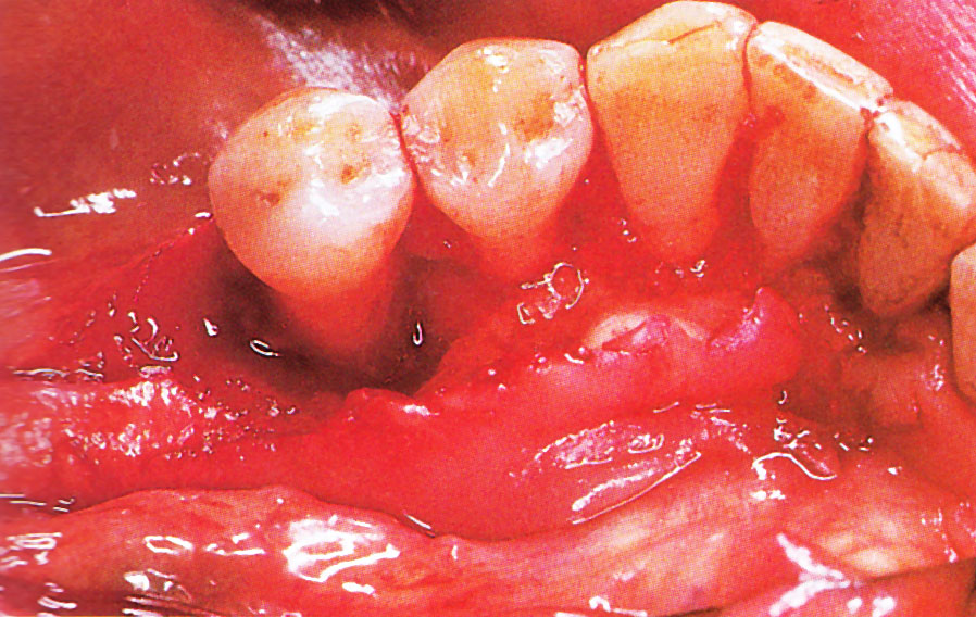
Prolonged bleeding: The dentist has a variety of means at their disposal to address bleeding; however, it is important to note that small amounts of blood mixed in the saliva after extractions are normal, even up to 72 hours after extraction. Usually, however, bleeding will almost completely stop within eight hours of the surgery, with only minuscule amounts of blood mixed with saliva coming from the wound. A gauze compress will significantly reduce bleeding over a period of a few hours.

Swelling: Often dictated by the amount of surgery performed to extract a tooth (e.g. surgical insult to the tissues both hard and soft surrounding a tooth). Generally, when a surgical flap must be elevated (i.e. and the periosteum covering the bone is thus injured), minor to moderate swelling will occur. A poorly-cut soft tissue flap, for instance, where the periosteum is torn off rather than cleanly elevated off the underlying bone, will often increase such swelling. Similarly, when bone must be removed using a drill, more swelling is likely to occur.

Sinus exposure and oral-antral communication: This can occur when extracting upper molars (and in some patients, upper premolars). The maxillary sinus sits right above the roots of maxillary molars and premolars. There is a bony floor of the sinus dividing the tooth socket from the sinus itself. This bone can range from thick to thin from tooth to tooth from patient to patient. In some cases it is absent and the root is in fact in the sinus. At other times, this bone may be removed with the tooth, or may be perforated during surgical extractions. The doctor typically mentions this risk to patients, based on evaluation of radiographs showing the relationship of the tooth to the sinus. It is important to note that the sinus cavity is lined with a membrane called the Sniderian membrane, which may or may not be perforated. If this membrane is exposed after an extraction, but remains intact, a “sinus exposed” has occurred. If the membrane is perforated, however, it is a “sinus communication”. These two conditions are treated differently. In the event of a sinus communication, the dentist may decide to let it heal on its own or may need to surgically obtain primary closure—depending on the size of the exposure as well as the likelihood of the patient to heal. In both cases, a resorbable material called “gelfoam” is typically placed in the extraction site to promote clotting and serve as a framework for granulation tissue to accumulate. Patients are typically provided with prescriptions for antibiotics that cover sinus bacterial flora, decongestants, as well as careful instructions to follow during the healing period.

Nerve injury: This is primarily an issue with extraction of third molars, but can occur with the extraction of any tooth should the nerve be close to the surgical site. Two nerves are typically of concern, and are found in duplicate (one left and one right): 1. the inferior alveolar nerve, which enters the mandible at the mandibular foramen and exits the mandible at the sides of the chin from the mental foramen. This nerve supplies sensation to the lower teeth on the right or left half of the dental arch, as well as sense of touch to the right or left half of the chin and lower lip. 2. The lingual nerve (one right and one left), which branches off the mandibular branches of the trigeminal nerve and courses just inside the jaw bone, entering the tongue and supplying sense of touch and taste to the right and left half of the anterior 2/3 of the tongue as well as the lingual gingiva (i.e. the gums on the inside surface of the dental arch). Such injuries can occur while lifting teeth (typically the inferior alveolar), but are most commonly caused by inadvertent damage with a surgical drill. Such injuries are rare and are usually temporary, but depending on the type of injury (i.e. Seddon classification: neuropraxia, axonotmesis, & neurotmesis), can be prolonged or even permanent.

Displacement of tooth or part of tooth into the maxillary sinus (upper teeth only). In such cases, almost always the tooth or tooth fragment must be retrieved. In some cases, the sinus cavity can be irrigated with saline (antral lavage) and the tooth fragment may be brought back to the site of the opening through which it entered the sinus, and may be retrievable. At other times, a window must be made into the sinus in the Canine fossa–a procedure referred to as “Caldwell luc”.

Dry socket (Alveolar osteitis) is a painful phenomenon that most commonly occurs a few days following the removal of mandibular (lower) wisdom teeth. It is commonly believed that it occurs because the blood clot within the healing tooth extraction site is disrupted. More likely,alveolar osteitis is a phenomenon of painful inflammation within the empty tooth socket because of the relatively poor blood supply to this area of the mandible (which explains why dry socket is usually not experienced in other parts of the jaws). Inflamed alveolar bone, unprotected and exposed to the oral environment after tooth extraction, can become packed with food and debris. A dry socket typically causes a sharp and sudden increase in pain commencing 2–5 days following the extraction of a mandibular molar, most commonly the third molar. This is often extremely unpleasant for the patient; the only symptom of dry socket is pain, which often radiates up and down the head and neck. A dry socket is not an infection, and is not directly associated with swelling because it occurs entirely within bone — it is a phenomenon of inflammation within the bony lining of an empty tooth socket. Because dry socket is not an infection, the use of antibiotics has no effect on its rate of occurrence. The risk factor for alveolar osteitis can dramatically increase with smoking after an extraction.

Bone fragments Particularly when extraction of molars is involved, it is not uncommon for the bones which formerly supported the tooth to shift and in some cases to erupt through the gums, presenting protruding sharp edges which can irritate the tongue and cause discomfort. This is distinguished from a similar phenomena where broken fragments of bone or tooth left over from the extraction can also protrude through the gums. In the latter case, the fragments will usually work their way out on their own. In the former case, the protrusions can either be snipped off by the dentist, or eventually the exposed bone will erode away on its own.


20/Feb/2011


What is Electrosurgery?

Electrosurgery is the application of a high-frequency electric current to biological tissue as a means to cut, coagulate, desiccate, or fulgurate tissue. (These terms are used in specific ways for this methodology—see below). Its benefits include the ability to make precise cuts with limited blood loss. Electrosurgical devices are frequently used during surgical operations helping to prevent blood loss in hospital operating rooms or in outpatient procedures.

In electrosurgical procedures, the tissue is heated by an electric current. Although electrical devices may be used for the cauterization of tissue in some applications, electrosurgery is usually used to refer to a quite different method than electrocautery. The latter uses heat conduction from a probe heated to a glowing temperature by a direct current (much in the manner of a soldering iron). This may be accomplished by direct current from dry-cells in a penlight-type device. Electrosurgery, by contrast, uses alternating current to directly heat the tissue itself. When this results in destruction of small blood vessels and halting of bleeding, it is technically a process of electrocoagulation, although “electrocautery” is sometimes loosely and nontechnically used to describe it. (Source from Wiakipidia)

Electrosurgery unit

Using electrosurgery in oral surgery procedures

Electrocautery is a very useful tool to make a cut or excise soft tissue just like a scaple blade does. While a cut is made; at the same time, electrosurgery coagulate the surrounding blood vessels make surgery bloodless. This will improve visibility during surgery and less blood loss. More over, healing of a wound with electrosurgery is proven to be faster that wound made by scaple blade.

Electrosurgery: Coagulating the gum area before impression taking during tooth preparation for crown fabrication

Dental procedure which can be done with

  • Excision of  lesions (eg. cysts, tumors)
  • Gum surgery
  • Implant placement
  • Crown lengthening
  • Coagulating the gum area before impression taking during tooth preparation for crowns/bridges

Advantages of using electrocautery

  • Less bleeding
  • Can be use to control bleeding
  • Wound heal faster with electrosurgery than using scaple blade


20/Feb/2011

In Malaysia, 2 persons are diagnosed with oral cancer every day where 75% seek treatment at the later stages and only half of them survives. According to the National Cancer Registry, Ministry of Health, mouth cancer is the third most common cancer among Malaysian Indian community. If cancer is detected earlier, the treatment outcome is more favorable and the chance of survival is definitely much higher. And of course, prevention is better than cure. Biopsy can be performed to detect the abnormalities of the swelling whether it is benign (good) or cancerous.

Tissue sample for biopsy


What is biopsy?


A biopsy is a procedure where a small part of tissue is removed so that it can be looked closely under microscope.

When is it indicated?

  • A sore or lesion on any area of your mouth which lasts for more than 2 weeks.
  • A white or red patch on your mouth.
  • An non-healing ulcer for more than 2 weeks.


What is the purpose of a biopsy?

a. For definitive diagnosis so that correct treatment can be initiated as soon as possible

b. To establish prognosis of a pre-cancerous or cancerous lesion


Types of biopsy

There are few types of biopsy but most commonly used are:

a. Incisional biopsy

  • A small portion of suspicious swelling will be removed and sent to the laboratory for diagnosis purpose.
  • Pathologists will determine the nature of the swelling and staging of swelling if it is found to be cancerous.
  • Often indicated when the swelling is large and has differing charecteristics, so it may need investigation on several areas.

b. Excisional biopsy

  • Whole swelling will be removed for both diagnosis and treatment purpose.
  • Often indicated when the swelling is small, usually less than 2cm.


How is it performed?

Excisional Biopsy

The procedure is painless as it is done under local anesthetic (numbing injection). Dentist will take small piece of the tissue and stitching is often required afterwards. The tissue specimen will then be labeled and stored into container with special solution inside. It will be sent to the laboratory for investigation. The procedure usually takes 15 minutes from start to finish.


So, if you have a persistent swelling inside your mouth, do seek a dentist for consultation. Don’t be panic, not all the swelling are cancerous but it is better to get it check as soon as possible.

Read More…


20/Feb/2011

Complex Extraction

Unfortunately, not all extractions can be done by simply grasping the tooth with forceps and rocking it out.  What if there is nothing left above the gum line to grasp? Or what if the crown breaks off leaving the roots still in the bone? These things can and do happen, and any dentist that extracts teeth will have to deal with them routinely.

Retain Root

In these cases, it becomes necessary to surgically remove the tooth.    This is frequently accomplished by prying the root out using a sharp instrument that can be forced between the root and the bone surrounding it.  This technique is called “luxation“.  In the case of multiple rooted teeth, the roots are first separated so they can be removed individually.  Unfortunately, not all roots or root fragments may be removed in this fashion. This means that the dentist must make an incision into the gums around the tooth and raise a flap of tissue exposing the tooth and its surrounding bone.

Surgical Extraction – a. Gum flap is raised and surrounding bone is removed. b. The roots of the molar are split with a drill  and removed. c. The flap is held  back in place with sutures

Sometimes, after the flap is raised, there is enough tooth exposed to grab and remove it as in a simple extraction.  Sometimes, the technique described above as luxation may successfully remove the tooth.  If luxation fails,  the dentist must take a handpiece (drill) and cut away some of the surrounding bone in order to gain a purchase on the tooth. After the tooth has been pried out of the artificially enlarged socket, the dentist then sutures (sews) the flap of tissue back in place so that healing can proceed normally.

Read more….


Get in touch

Contact us now

Book an appointment with our doctors now!! Click here
If you have any question, don’t hesitate to contact us, we are more than glad to provide you with the information you need!

We accept Cash, Credit Card, Grabpay, Alipay, Touch n Go, MayBank QRPAY and Boost


Follow us

Our Activity

Curious about what we do every day? Follow us to learn more about the dental health care we provide.


Copyright 2023. All rights reserved.