Lateral cephalometric is a radiograph of the head taken with the x-ray beam perpendicular to the patient’s sagittal plane. This radiography image is useful for studying the dental and facial growth of a patient.

In orthodontic treatment, clinician/orthodontist use this radiograph to identified dental and skeletal deformity; the relationship between upper and lower teeth and jaw bone. By doing so, he can create a treatment plan to correct the teeth misalignment. This radiograph is also useful in monitoring the progress of braces treatment and to compare before and after treatment.

During radiograph taking, the patient’s head is positioned in the most relax position — natural head position — is a standardized orientation of the head that is reproducible for each individual and is used as a means of standardization during analysis of dentofacial morphology both for photos and radiographs.

Cephalometric Tracing
Cephalometric tracing is an overlay drawing produced from a cephalometric radiograph by digital means and a computer program or by copying specific outlines from it with a lead pencil onto acetate paper, using an illuminated view-box. Tracings are used to facilitate cephalometric analysis, as well as in superimpositions, to evaluate treatment and growth changes.
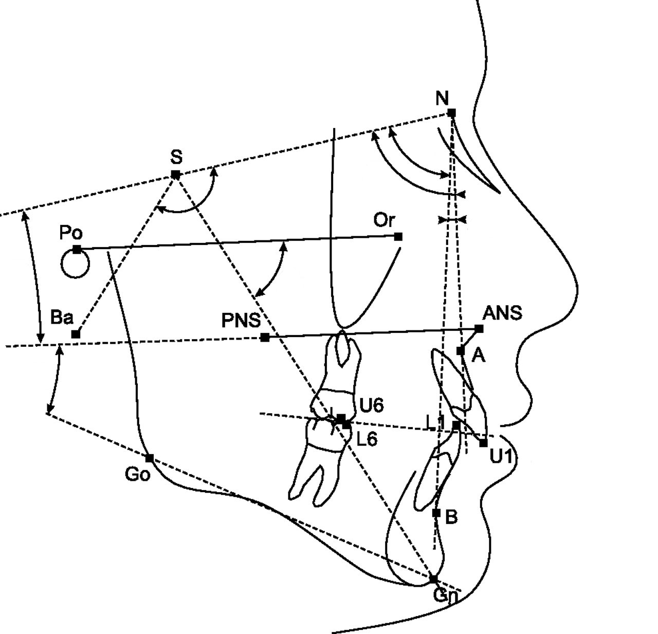
The landmarks on the radiograph are identified and marked.

Lines are made to join the markings and the angles where the lines intercept will be recorded. The values of the angle will be compared to the norm value according to the patient’s race. And finally, a conclusion will be depends on the deviation of the patient’s value to the norm value.
Sometimes, lateral cephalometric radiograph can be use to compare before and after treatment or the growth pattern of a child. For example, in the image below, the green line represent before treatment and the black will be after treatment. From here, we can appreciate how the teeth and jaws move during treatment. Doctor use this radiograph to evaluate if he has achieve his treatment objective!!

Other Radiograph Modalities…
- CBCT (Cone Beam CT)
- Lateral Cephalometry Radiograph
- Dental Panoramic Tomogram (OPG)
- Intra-oral Radiograph
 :
:  :
: 