BlogNEWS AND DENTAL EDUCATION
We share informative articles and news.

What is an apex locator?

An apex locator (or electronic apex locator – EAL) is an electronic device used in endodontics/root canal therapy to determine the position of the apical foramen and thus determine the length of the root canal space. The apex of the root has a specific resistance to electrical current, and this is measured using a pair of electrodes typically hooked into the lip and attached to an endodontic file. The electronic principle is relatively simple and is based on electrical resistance; when a circuit is complete (tissue is contacted by the tip of the file), resistance decreases markedly and current suddenly begins to flow. According to the device, this event is signaled by a beep, a buzz, a flashing light, digital readouts, or a pointer on a dial. – source from wikipedia
Role of Electronic Apex/ Foramen Locators
Working length determination during root canal therapy has been greatly aided by Electronic apex locators (EALs). They can save time in determining initial working length, save on the number of X rays taken, and can be particularly helpful when the periapex is unclear on radiographs or when there is a question about whether a perforation has occurred.
As the matter of fact, foramen does not coincide with the anatomical apex in most teeth. You can confirm this fact by examining the morphology of extracted teeth. One of the earliest brands was Root ZX by JMorita of Japan which still is the gold standard in accuracy as confirmed by various studies. We have been using it for many years. Intraoral radiographs do not show the apical foramen and are often misleading, leading to compromising results, see below. Picture shows how radiographs can be deceptive in determing correct working length-WL, you will never be able to assess correct WL without a good apex locator.
Root ZX II – Accurate apical foramen measurement

Currently, we are using Root ZX apex locator for our root canal treatments.
Invented in 1992, the J. Morita Root ZX II is currently one of the best selling apex locators. It has proven itself safe and accurate, and was the first unit providing dentists the capability to work in wet canals. Blood and other fluids do not typically affect measurements.
Root ZX II patented technology offers an accuracy rate above 96%. LCD readout is large and easy to read. Action of the meter in the display corresponds to the tactile sensation of using the file.
Other Root ZX II Features:
- slim, lightweight file holder
- no zero-adjustment
- automatic calibration
- battery power indication
- automatic power off function
An in vitro comparison of three apex locators concluded that the Root ZX II was significantly more accurate at finding the apical foramen than competitive models.
Read more
 An articulator is a mechanical device used in dentistry which represents the anatomy of temporomandibular joint (the joint connecting lower jaw to the skull), upper jaw and lower jaw of patient to which upper teeth cast and lower teeth cast are fixed to the articulator in order to reproduce patient’s jaw movements.
By nature, the purpose of these articulators can only be achieved when the position of the maxilla is duplicated with respect to the skull. The upper teeth cast should be mounted on a semi-adjustable articulator using a face bow. The closer the articulator matches the patient’s anatomy, the better the treatment outcome will be, hence shorter dental treatment time is required.
It is a complex articulator which almost imitates the anatomy of the temporomandibular joint and follows the movement of your lower jaw. Therefore, it can be used in the fabrication of complex crowns, long span bridges and full mouth rehabilitation. This articulator is also used for jaw surgery (orthognathic) planning.
Semi-adjustable articulator is adjustable in certain areas but not all. They have adjustable horizontal condylar paths, adjustable lateral condylar paths, adjustable incisal guide tables, and adjustable intercondylar distances. Nevertheless, this articulator is adequate for most of the cases.
An articulator is a mechanical device used in dentistry which represents the anatomy of temporomandibular joint (the joint connecting lower jaw to the skull), upper jaw and lower jaw of patient to which upper teeth cast and lower teeth cast are fixed to the articulator in order to reproduce patient’s jaw movements.
By nature, the purpose of these articulators can only be achieved when the position of the maxilla is duplicated with respect to the skull. The upper teeth cast should be mounted on a semi-adjustable articulator using a face bow. The closer the articulator matches the patient’s anatomy, the better the treatment outcome will be, hence shorter dental treatment time is required.
It is a complex articulator which almost imitates the anatomy of the temporomandibular joint and follows the movement of your lower jaw. Therefore, it can be used in the fabrication of complex crowns, long span bridges and full mouth rehabilitation. This articulator is also used for jaw surgery (orthognathic) planning.
Semi-adjustable articulator is adjustable in certain areas but not all. They have adjustable horizontal condylar paths, adjustable lateral condylar paths, adjustable incisal guide tables, and adjustable intercondylar distances. Nevertheless, this articulator is adequate for most of the cases.
 Uses of articulators
- Educate to patient their jaw relation
- Reproduce patient’s jaw movement like opening and closing of mouth
- To diagnosis the state of patient’s occlusion
- To help in treatment planning
- To help in fabrication and modifying of dental restoration (dental crown and bridge)
- To obtain good occlusion for dental restoration
- For jaw surgery planning
- Allows teeth arrangement for denture
- Dentist allows to adjust patient’s dental restoration on the articulator (outside patient’s mouth) without the disturbance of patient’s tongue, cheek, and saliva.
- Reduce dental visits and treatment time because articulator helps to resemble patient’s jaw relation.
- Adjustment and correction of dental restoration can be done in the absence of patient
- With the help of articulator, your dentist allows to visualize the inner side of your teeth easily
…
“Why does my dentist takes photo of my teeth?”
I’m sure some of you have been wondering about it. So here’s why dentist takes photo of your teeth and even portraits sometimes.

- Photography act as a vital communicating tool between dentist and the patient so that a clearer message or explanation can be delivered.
- Imperfections that are not readily visible to the patient will be apparent in still photographs, patient get to visualise his/her oral condition.
- Establish a baseline so that dentist able to monitor any recession or suspicious lesions that you may have in your mouth to discover if these conditions are getting better or worse.
- Document cases by dentist for recording purpose.
DSLR Camera
Multiple system can be choose for dental photography and generally work well for practices. However, the digital single lens reflex (DSLR) camera still the most ideally suited for practices that to use photography for documentation for lectures or publications.
Basic kit for dental photography:
- DSLR camera system
- Macro lens (85–105 mm)
- External ring flash / Dual Flash
- Retractors
- Mirror
- Contrastor
-

Digital SLR Camera


Intra-Oral Photos
Multiple views of intra-oral photos will be taken. The basic views are:
- Front view without retractor
- Front view with retractor
- Left buccal view
- Right buccal view
- Upper occlusal view
- Lower occlusal view

Dentist might take some extra photos which specifically focus on a tooth/ area to focus on the problematic part.
Studio Photography
Sometimes additional extra-oral photos are needed. In our clinic, there’s a room set up with some additional tools for portraits such as NiceFoto TB-400C Flash with a diffuser/ softbox, and a white lighting board mounted on a wall.

Extra Oral Photos
For extra-oral, pictures of patient must include full face. The basic views are:
- Front profile view
- Front smiling view
- Left & Right profile view
- Left & Right 45° profile view

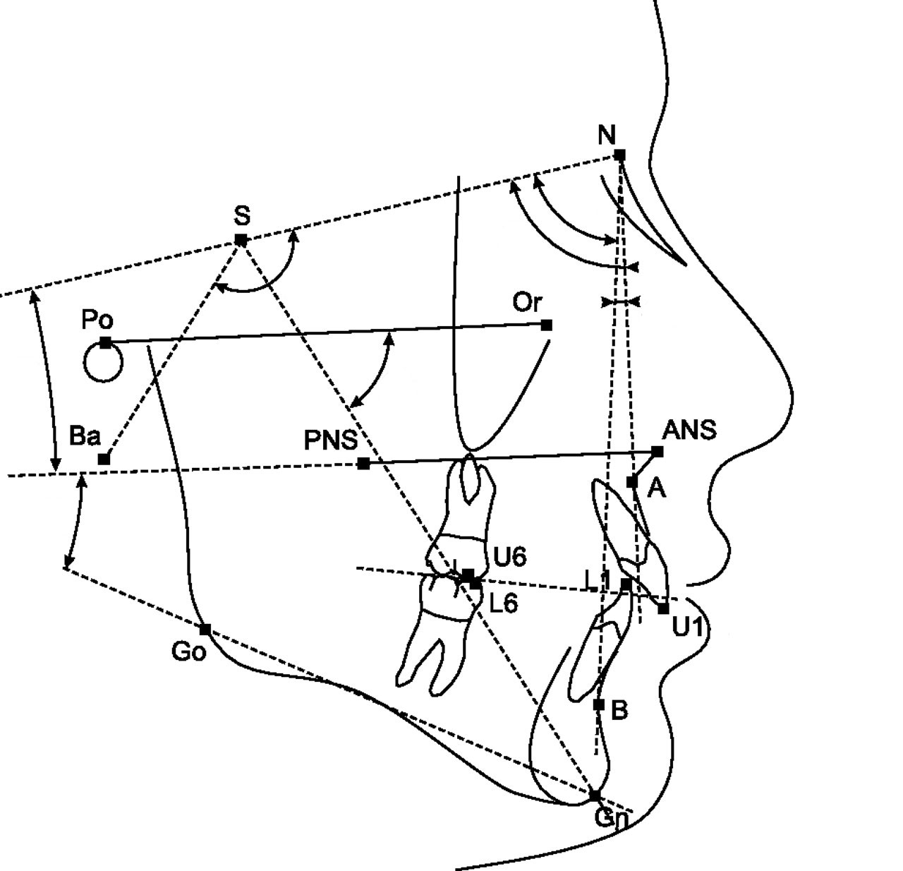
Lateral cephalometric is a radiograph of the head taken with the x-ray beam perpendicular to the patient’s sagittal plane. This radiography image is useful for studying the dental and facial growth of a patient.

In orthodontic treatment, clinician/orthodontist use this radiograph to identified dental and skeletal deformity; the relationship between upper and lower teeth and jaw bone. By doing so, he can create a treatment plan to correct the teeth misalignment. This radiograph is also useful in monitoring the progress of braces treatment and to compare before and after treatment.

During radiograph taking, the patient’s head is positioned in the most relax position — natural head position — is a standardized orientation of the head that is reproducible for each individual and is used as a means of standardization during analysis of dentofacial morphology both for photos and radiographs.

Cephalometric Tracing
Cephalometric tracing is an overlay drawing produced from a cephalometric radiograph by digital means and a computer program or by copying specific outlines from it with a lead pencil onto acetate paper, using an illuminated view-box. Tracings are used to facilitate cephalometric analysis, as well as in superimpositions, to evaluate treatment and growth changes.
The landmarks on the radiograph are identified and marked.

Lines are made to join the markings and the angles where the lines intercept will be recorded. The values of the angle will be compared to the norm value according to the patient’s race. And finally, a conclusion will be depends on the deviation of the patient’s value to the norm value.
Sometimes, lateral cephalometric radiograph can be use to compare before and after treatment or the growth pattern of a child. For example, in the image below, the green line represent before treatment and the black will be after treatment. From here, we can appreciate how the teeth and jaws move during treatment. Doctor use this radiograph to evaluate if he has achieve his treatment objective!!

Other Radiograph Modalities…
- CBCT (Cone Beam CT)
- Lateral Cephalometry Radiograph
- Dental Panoramic Tomogram (OPG)
- Intra-oral Radiograph
Dental Panoramic Tomogram or OPG is a sophisticated x-ray machine used to take radiographic images of the teeth and jaws bone which is in a arch position. We used to call this machine as OPG (which stands for ORTHOPANTOGRAM), which was named after the first x-ray unit.
OPG performed by using a technique called “tomography”.
The X-ray tube moves around the head, the x-ray film moves in the opposite direction behind your head. This generates an image slice where the mandible and teeth are in focus, and the other structures are blurred.


Anatomy
The anatomy consists of the body, ramus and angle of mandible, coronoid process, mandibular notch, condyle of mandible, alveolar ridge, symphysis menti, maxillary sinuses, nasal fossae and 16 upper and 16 lower teeth, as shown in the image below.

Reasons for OPG requests
Dental Disease
- Caries – appear as different shaped areas of radiolucency in the crowns or necks of teeth.
- Peridontioiditis – when inflammation extends into the underlying alveolar bone and there is a loss of attachment.
- Peridontal Abscess – Radiolucent area surrounding the roots of the teeth.
Impacted or embedded teeth (eg. wisdom teeth)
- OPG shows angulation, shape of roots, size and shape of crown, effect on other teeth.
- To look for impacted canine
Teeth Abnormalities
- Eg. Developmental, to show size, number, shape and position.
Lesions in the jaw bone
- Cyst of jaw bone – shape, size, extension, involving nearby structure
- Tumour/growth – benign, malignant
Trauma to teeth and facial skeleton
- Mandible fractures are often bilateral.
- Panoramic view of mandible to view the fracture.
- Determine site and direction of fracture lines.
- Relationship of teeth to fracture lines.
- Alignment of bone fragments after healing.
- Evidence of infection or other complications post intervention.
- Follow up to assess healing.
Planing for implant placement
- To identify the position and location for implant placement
- Bone quality and quantity
- Anatomical structure that should be avoided such as the maxillary sinus and the inferior dental nerve
Other Radiograph Modalities…
- CBCT (Cone Beam CT)
- Lateral Cephalometry Radiograph
- Dental Panoramic Tomogram (OPG)
- Intra-oral Radiograph
Dental intra-oral radiographs a.k.a X-rays. Dentists use radiographs for multiple reasons: to find hidden dental structures, malignant or benign masses, bone loss, and cavities.
How does x-ray of your teeth formed?
X-ray of your teeth is formed by a controlled burst of X-ray radiation which penetrates oral structures at different levels, depending on varying anatomical densities, before striking the film or sensor.
- Teeth appear lighter because less radiation penetrates them to reach the film.
- Dental caries, infections and other changes in the bone density, and the periodontal ligament, appear darker because X-rays readily penetrate these less dense structures.
- Dental restorations (fillings, crowns) may appear lighter or darker, depending on the density of the material.
Should Patients Have Concerns About Radiation Exposure?
The dosage of X-ray radiation for dental is typically safe, around 0.150 mSv for a full mouth series, according to the American Dental Association website. It is equivalent to a few days’ worth of background environmental radiation exposure, or similar to the dose received during a cross-country airplane flight (concentrated into one short burst aimed at a small area).
Incidental exposure is further reduced by the use of a lead shield, lead apron, sometimes with a lead thyroid collar. Operator exposure is reduced by stepping out of the room, or behind adequate shielding material, when the X-ray source is activated.
Types of intra-oral radiographs
- Bitewing
- Periapical
Bitewing radiograph

Bitewing radiograph designed the placement of the film packet to reveal the coronal halves of the maxillary and mandibular teeth, inter-proximal contacts and portions of the interdental septa.
It is indicated primarily to detect or monitor interproximal caries if the proximal surfaces of the teeth cannot be visually or tactilely examined.
Occlusal caries, crestal alveolar bone level and secondarily for eruption patterns, caries and restoration proximity to pulp spaces, primary molar furcation pathology and developmental anomalies may also be detected with bitewing radiographs.
Periapical Radiograph
PA radiography describes intra-oral techniques designed to show individual teeth and the tissues around the apices. Each image usually shows two to four teeth and provides detailed information about the teeth and the surrounding alveolar bone.


Indications for PA radiograph are:
- Detection of apical infection/inflammation
- Assessment of the periodontal status
- After trauma to the teeth and associated alveolar bone
- Assessment of the presence and position of unerupted teeth
- Assessment of root morphology before extractions
- During endodontics
- Preoperative assessment and postoperative appraisal of apical surgery
- Detailed evaluation of apical cysts and other lesions within the alveolar bone
- Evaluation of implants postoperatively.
Other Radiograph Modalities…
- CBCT (Cone Beam CT)
- Lateral Cephalometry Radiograph
- Dental Panoramic Tomogram (OPG)
- Intra-oral Radiograph
Sterilization
Dental instrument that are used or contaminated have to be cleaned and bacteria-free before reuse. Therefore, they need to be sterilized before use. Sterilization is a term referring to any process that eliminates (removes) or kills all forms of life, including transmissible agents (such as fungi, bacteria, viruses, spore forms, etc.) present on a surface, contained in a fluid, in medication, or in a compound such as biological culture media. Sterilization can be achieved by applying the proper combination of heat, chemicals, irradiation, high pressure, and filtration. (Source from wikipedia)

Autoclave
In dentistry, we use autoclave to sterilize our dental instruments. Autoclave is a device to sterilize equipment and supplies by subjecting them to high pressure saturated steam at 121 °C or more, typically for 15–20 minutes depending on the size of the load and the contents. It was invented by Charles Chamberland in 1879, although a precursor known as the steam digester was created by Denis Papin in 1679. The name comes from Greek auto, ultimately meaning self, and Latin clavis meaning key — a self-locking device. (Source from wikipedia)
Most dental clinic use autoclave unit to sterilize their instruments. According to the European Standard EN 13060, autoclave are divided into:

Type B- It has 3-times per-vacuum preceding vacuum drying. It can be used on wrapped and hollow instruments, which means a piece of equipment can be sterilized now for use later. This is the most effective autoclave as the steam able to penetrates deep into the pouches/wrappers or even double pouched instruments.
Type S – Comes with a one times pre-vacuum and vacuum drying function and efficient quick spraying steam generator. It can’t be used to sterilize instruments which are double pouch or the instruments which are wrapped in the thick wrapper/pouch.
Type N – This autoclave comes without vacuum function, it can be used for hollow instruments and solid instruments.This autoclaves are only suitable for a specific type of
load–for solid, unwrapped instruments.

Type B European Standard Autoclave – It has the highest standard among the type S and type N. It allows deep penetration into pouched/wrapped instrument. Type B Autoclave used widely in operating theater and it is used by our clinic too.
Book an appointment with our doctors now!! Click here
We accept Cash, Credit Card, Grabpay, Alipay, Touch n Go, MayBank QRPAY and Boost
Copyright 2023. All rights reserved.
Get in touchContact us now
If you have any question, don’t hesitate to contact us, we are more than glad to provide you with the information you need!
 :
:  :
: 